Many recent achievements of IMIC laboratory have been accepted by top conferences in the field.
- IMIC Lab
- Research , Research results
- June 25, 2026
From top journals to top conferences, the academic influence of Jiangsu University Key Laboratory of Intelligent Medical Image Computing (IMIC) is expanding in multiple dimensions. The IMIC Lab has been reporting frequently recently. A number of original results focusing on cutting-edge exploration have not only been steadily published in top journals, but have also successfully entered many top academic conferences. This article will introduce in detail the latest research progress of the IMIC laboratory at the top conference stage in the field.
Result 1: The paper of Liu Mingxin, a doctoral student in IMIC laboratory, was accepted by MICCAI 2026
Recently, the research paper “Synergistic Information Disentanglement for Omni-modal Slide Representation Learning in Computational Pathology” completed by Liu Mingxin (supervisor: Professor Xu Jun), a doctoral student in the IMIC laboratory, was accepted in advance (Top 9%) by the top international conference in the field of medical image computing, “The 29th International Conference on Medical Image Computing and Computer-Assisted Intervention (MICCAI 2026)”. MICCAI is one of the most influential top international conferences in the field of medical image computing and computer-assisted intervention. The conference will be held this year from September 27 to October 1 at the Congress Center in Strasbourg, France.
This study focuses on three long-term challenges faced by whole-slide image (WSI) modeling in computational pathology: insufficient utilization of multi-modal information, redundant alignment of modalities caused by traditional contrastive learning, and difficulty in effectively modeling cross-modal synergistic information. A full-modal slice representation learning framework Φ-Omni based on synergistic information decoupling theory is proposed. This method (shown in Figure 1) takes three modalities of pathology images, genome information, and pathology reports as input, and extracts WSI patch features, transcriptome expression features, and clinical text semantic features through modality-specific encoders. On this basis, the model designed a Synergistic Information Bottleneck (SIB) module, which reduces modal redundant noise while retaining cross-modal key information interactions through a layer-by-layer compressed embedding space structure, thereby forming a compact global joint representation. Furthermore, this study starts from the Partial Information Decomposition (PID) theory and proposes the Synergistic Information Disentanglement (ΦID) optimization goal, which decomposes multi-modal information into three types of components: redundant information, specific information and collaborative information, and explicitly maximizes the cross-modal non-decomposable collaborative information (Φ) while suppressing single-modal interpretable redundant information. By combining Gaussian Canonical Projector (GCP), this method stably estimates information entropy in a differentiable high-dimensional space and achieves optimal modeling of collaborative information. In the pre-training stage, the model simultaneously introduces contrastive learning alignment loss and ΦID collaborative decoupling loss. Through dual-objective joint optimization, the model can maintain cross-modal semantic consistency while avoiding the representation collapse problem caused by traditional contrastive learning, thus improving the expression and generalization capabilities of multi-modal representation.
The method was systematically validated on two TCGA large-scale cohorts of breast cancer and non-small cell lung cancer, and downstream task evaluation was carried out on five independent external data sets including BRACS, CPTAC-BRCA, and CPTAC-NSCLC. Under the few-shot learning setting (k=1/5/10/25), Φ-Omni achieves leading performance in multiple classification tasks. For example, in the BRACS breast cancer classification task, the AUC reaches 83.3% when k=25; in the CPTAC-NSCLC data set, the AUC reaches 99.4% when k=25. The overall performance is better than existing methods including TANGLE, CLAM, TransMIL and various pathological basic models. Ablation experiments further show that the Synergistic Information Bottleneck (SIB) and ΦID decoupling goals have a key contribution to model performance. The synergistic information maximization mechanism can significantly improve the quality of cross-modal representation, while the redundant compression mechanism effectively suppresses the modal collapse phenomenon, allowing the model to show stronger stability and generalization capabilities under different tasks and data distributions.
This research achieves efficient few-shot transfer learning capabilities for WSI without the need for additional task fine-tuning, and significantly improves the modeling capabilities of multi-modal pathological AI models for complex biomedical information. Relevant results provide a new theoretical framework and method path for full-modal computational pathology learning, and also provide an important reference for the application of cross-modal collaborative modeling in medical artificial intelligence. In the future, this method is expected to be further expanded to scenarios such as cross-center data fusion, weakly supervised pathological diagnosis, and multi-omics integrated analysis, providing more reliable technical support for precision medicine and intelligent diagnosis.
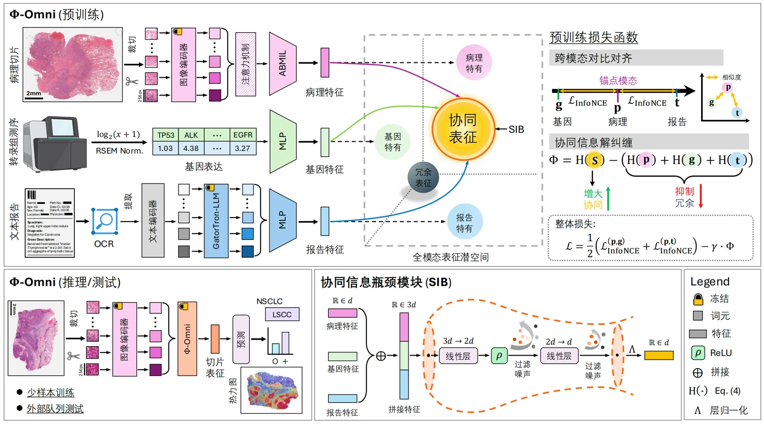
Figure 1. Schematic diagram of the framework of the full-modal slice representation learning method based on collaborative information decoupling proposed in this article
Paper information
Mingxin Liu, Chengfei Cai, Anwen Lu, Pengbo Xu, Jun Li, Jinze Li, Depin Chen, Jun Xu, “Synergistic Information Disentanglement for Omni-modal Slide Representation Learning in Computational Pathology,” In International Conference on Medical Image Computing and Computer-Assisted Intervention (MICCAI), 2026.
Result 2: The paper of Associate Professor Luo Yuemei’s research group in IMIC Lab was accepted by IEEE ICASSP 2026
Recently, the paper “Automatic Identification of Retinopathy from Optical Coherence Tomography Images via a Similarity Matching Approach” by the research group of Associate Professor Luo Yuemei of the IMIC Lab was accepted by the 2026 IEEE International Conference on Acoustics, Speech, and Signal Processing (IEEE ICASSP 2026). ICASSP is the oldest and top international conference of the IEEE Signal Processing Society. The 51st ICASSP will be held in Barcelona, Spain, from May 4 to 8, 2026.
This study focuses on issues such as the high cost of annotating clinical OCT images, the scarcity of professional annotated samples, and the vulnerability of traditional semi-supervised methods to low-quality pseudo-labels. It proposes a semi-supervised retinopathy recognition method that simultaneously integrates semantic-level and instance-level consistency. The model (shown in Figure 2) uses pre-trained Vision Transformer (ViT) as the backbone network: for labeled OCT images, supervised learning is completed through weakly enhanced samples and real labels; for unlabeled images, weakly enhanced and strongly enhanced views are constructed, pseudo labels are generated and filtered at the semantic prediction level, and instance similarities are calculated in the feature space. Furthermore, this method uses expansion and aggregation operations to calibrate and interactively update semantic pseudo-labels and instance pseudo-labels, and jointly optimizes supervised loss, semantic-level unsupervised loss, and instance-level consistency loss, thereby improving the reliability of pseudo-labels and the classification robustness of the model under very few labeling conditions.
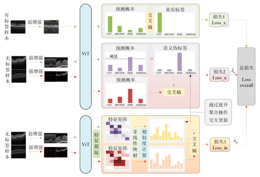
Figure 2. Schematic diagram of the semi-supervised OCT classification framework based on similarity matching proposed in this article
The method was systematically validated on two public OCT datasets, BOE and CELL. In the BOE data set, only 55 annotated images (accounting for 1.70% of all samples) are used for training, and the classification accuracy reaches 96.4%, and the AUC reaches 99.7%; in the CELL data set, only 80 annotated images (accounting for 0.095% of all samples) are used, the classification accuracy reaches 97.0%, and the AUC reaches 99.5%. Compared with various traditional supervised learning and mainstream semi-supervised learning methods, the accuracy of the proposed method on the two data sets is improved by 2.2 and 3.4 percentage points respectively. Ablation experiments further show that semantic-level consistency and instance-level similarity learning can complement each other, and the complete model achieves the best performance.
This study can still achieve stable and accurate OCT retinopathy recognition at a very low annotation ratio, significantly reducing the reliance of medical image intelligent analysis on large-scale expert annotation data. Relevant results provide new technical paths for automatic screening and auxiliary diagnosis of retinal diseases in scenarios with limited annotation resources, and also provide valuable research ideas for semi-supervised medical image learning driven by semantic information and instance relationships. In the future, this method can be further expanded to cross-device, cross-center and multi-modal ophthalmic image analysis, providing more reliable artificial intelligence technology support for early screening of eye diseases and clinical decision support.
Paper information
Yuemei Luo, Yuan Li, Jiaxue Mei, Jun Xu, Linbo Liu, and Xiaohua Qian, “Automatic Identification of Retinopathy from Optical Coherence Tomography Images via a Similarity Matching Approach,” In ICASSP 2026-2026 IEEE International Conference on Acoustics, Speech and Signal Processing (ICASSP) , pp. 8072-8076, 2026.
Achievements 3: The paper of Dr. Ming Wenlong’s research group from IMIC Lab was accepted by MIDL 2026
Recently, the research paper “D²-Former: Mixture-Of-Experts Guided Dual Transformer for Multi-Scale Medical Image Segmentation (D²-Former: Mixture-Of-Experts Guided Dual Transformer for Multi-Scale Medical Image Segmentation)” by the research group of Professor Ming Wenlong of IMIC Lab was accepted by the International Conference on Medical Imaging with Deep Learning (MIDL 2026). MIDL is an emerging top international conference at the intersection of medical imaging and deep learning, focusing on cutting-edge research of deep learning methods in medical image analysis, computer-aided diagnosis, disease screening, prognosis assessment and treatment decision-making. The conference will be held in Taipei, China, from July 8 to 10 this year.
Medical image segmentation is a key task in computer-aided diagnosis and treatment planning. However, real clinical images often have problems such as blurred boundaries, large scale changes, and complex tissue morphology, making it difficult for the model to simultaneously take into account global semantic understanding and local detail recovery. In multi-modal, multi-structure and multi-scale scenarios, existing methods still face challenges such as insufficient adaptive feature fusion, insufficient fine-grained boundary characterization, and insufficient generalization capabilities. In response to the above problems, the research team proposed the D²-Former medical image segmentation framework (shown in Figure 3). This method uses a dual encoder-decoder structure to combine Swin Transformer with the DINOv3 basic model to extract hierarchical structural features and high-quality semantic representation respectively. At the same time, the model introduces the Softer Mixture-of-Experts (Softer-MoE) mechanism in Swin Transformer, which enables the network to adaptively refine features based on input content to better handle complex anatomical structures and fuzzy boundaries. In terms of feature fusion, the study designed the Spatial-Frequency Gated Channel Attention (SF-GCA) module to fuse the complementary information of the two coding branches through spatial alignment and gated attention mechanisms. Subsequently, the model uses Residual Attention Decoder (RAD) to gradually restore high-resolution segmentation results, and combines spatial and channel attention and deep supervision strategies to further improve boundary reconstruction capabilities and multi-scale prediction stability.
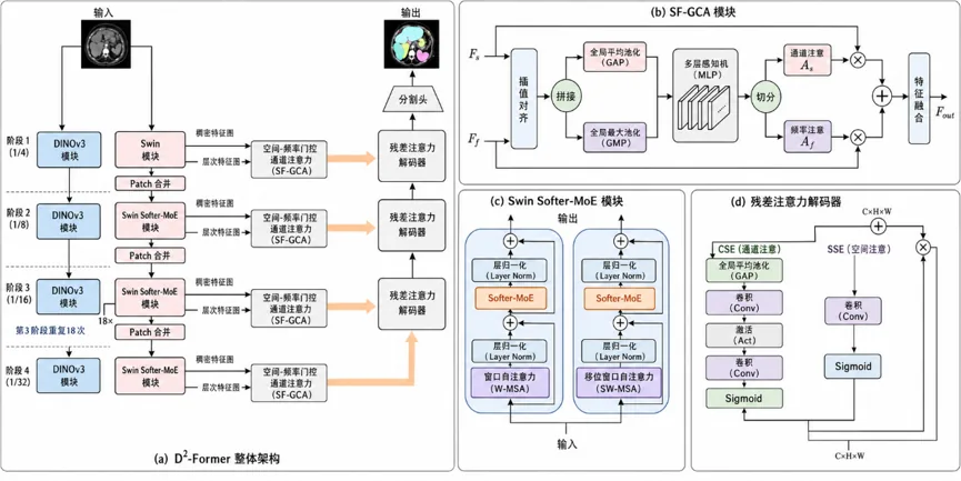
Figure 3. Schematic diagram of the D²-Former multi-scale medical image segmentation framework proposed in this article
The method was systematically verified on nine public medical image segmentation datasets, covering tasks such as polyp segmentation, retinal blood vessel segmentation, multi-organ abdominal CT segmentation, and pathological cell nucleus segmentation. On the Synapse multi-organ abdominal CT segmentation data set, D²-Former achieved an average Dice of 85.53% and an mIoU of 77.32%, which is better than a variety of representative methods; on the DRIVE and STARE retinal blood vessel segmentation data sets, it achieved an F1-score of 83.21% and 82.11% respectively, showing good recognition of small, low-contrast vascular structures. In addition, this method also achieved competitive results in polyp segmentation and pathological cell nucleus segmentation tasks, demonstrating good cross-task adaptability. Ablation experiments show that the DINOv3 branch, SF-GCA fusion module, RAD decoder and Softer-MoE mechanism all help improve model performance. Multiple independent training results further show that the method has smaller performance fluctuations under different random seeds and has better stability and reproducibility.
This research addresses issues such as scale changes, boundary blur, and cross-task generalization in medical image segmentation, and explores a collaborative modeling approach of basic visual models, hierarchical Transformers, and hybrid expert mechanisms. Relevant results provide new network design ideas for multi-scene medical image segmentation, and also provide reference for the application of basic model feature migration, dynamic expert routing and multi-scale feature fusion in medical image intelligent analysis.
Paper information
Md Sohag Mia, Aya Taourirte, Muhammad Abdullah Adnan, and Wenlong Ming, “D²-Former: Mixture-Of-Experts Guided Dual Transformer for Multi-Scale Medical Image Segmentation,” Medical Imaging with Deep Learning , 2026.
Achievements 4: The paper of Dr. Ming Wenlong’s research group from IMIC Lab was accepted by MICCAI 2026
Recently, the research paper “Noise-Aware Importance–Uncertainty Disentangled Multimodal Learning for Robust Cancer Survival Prediction” by the research group of Professor Ming Wenlong of the IMIC Lab was accepted by the 2026 International Conference on Medical Image Computing and Computer-Assisted Intervention (MICCAI 2026).
This study focuses on the key problem of modality-specific technical noise and the difficulty in collaborative modeling of heterogeneous prognostic features in multimodal cancer prognosis prediction, and proposes the importance of noise-awareness - Uncertainty Decoupled Multimodal Survival Network (NADMSurv) (as shown in Figure 4). The model takes whole-slice image (WSI) and mRNA expression profile as bimodal inputs: WSI has stable prognostic clues but limited discriminative power, while mRNA For modal asymmetry that has strong prognostic relevance but is severely interfered by technical noise, the method first jointly performs feature pre-screening and patient-level noise quantification through the importance and uncertainty dual-track module to capture heterogeneous prognostic patterns; then, the noise-aware decoupling mechanism is used to separate the multi-modal representation into shared representation and modality-specific representation, achieving explicit separation of cross-modal complementary information and modality-unique signals. In order to model high-order patient correlations, the method introduces a hypergraph neural network to construct group-level correlations (as shown in Figure 5), and combines subjective logic to convert risk scores into evidence opinions. Finally, evidence-level uncertainty fusion and robust prognosis prediction are achieved through cross-attention and consensus operators.
The method was systematically validated on five cancer cohorts from the TCGA database (breast invasive carcinoma BRCA, lung adenocarcinoma LUAD, gastric adenocarcinoma STAD, brain low-grade glioma LGG, and clear cell renal cell carcinoma KIRC). Compared with nine existing supervised, single-modal and multi-modal methods, NADMSurv achieves the best C-index performance in all five cohorts. In addition, Kaplan-Meier survival analysis based on model prediction risk stratification showed that the survival probabilities of patients in the high-risk group and the low-risk group were significantly different (log-rank test p values for all cohorts were less than 0.001), fully verifying its effectiveness in clinically actionable risk stratification.
This study establishes a new paradigm for multimodal cancer survival prediction under technical noise conditions by explicitly modeling the importance-uncertainty asymmetry between WSI and mRNA, improving the robustness and reliability of prognostic prediction. Relevant results provide a new technical path for precise oncology decision-making by integrating pathological images and genomics data, and also provide valuable research ideas for noise-aware multi-modal representation learning and evidence-level fusion mechanisms. In the future, this method can be further expanded to more cancer types, multi-center heterogeneous data and multi-omics integrated analysis, providing more reliable artificial intelligence technology support for personalized treatment decision-making and clinical prognosis assessment.
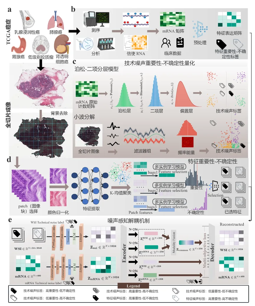
Figure 4. Schematic diagram of the NADMSurv model framework proposed in this article
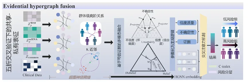
Figure 5. Schematic diagram of the evidence hypergraph fusion architecture proposed in this article
Paper information
Wenlong Ming, Wenbin Ye, Mingxin Liu, Depin Chen, Yiping Jiao, Jun Xu, and Xiangxue Wang, “Noise-Aware Importance–Uncertainty Disentangled Multimodal Learning for Robust Cancer Survival Prediction,” In MICCAI 2026: 29th International Conference on Medical Image Computing and Computer Assisted Intervention (MICCAI) , 2026.
Translated from the original Chinese source.