A collaborative paper between IMIC Lab and Fudan University Cancer Hospital was published in the collaborative journal npj Precision Oncology in Nature
- IMIC Lab
- News , Comprehensive news
- January 25, 2026
Recently, the multi-modal medical data analysis innovation team of Nanjing University of Information Science & Technology, Jiangsu University Key Laboratory of Intelligent Medical Image Computing (IMIC) and related teams of Fudan University Cancer Hospital collaborated and published in the collaborative journal npj Precision Oncology of Nature.
The study is titled “Interpretable Deep Learning Model Based on H&E Stained Sections for Predicting Endometrial Cancer Molecular Subtypes.” This research is dedicated to integrating advanced image analysis and deep machine learning technology based on conventional H&E stained sections to quantitatively extract histomorphological features and build a prediction model that can intelligently distinguish different molecular subtypes. The model predicts the four main molecular subtypes of endometrial cancer by analyzing conventional pathological H&E stained sections. Before the model is input, all full-field sections undergo a systematic preprocessing process: first, the sections are cut into fixed-size non-overlapping image blocks; then blank or over-stained invalid areas are excluded through brightness filtering; then the Vahadane method is used for staining normalization to reduce the impact of staining differences between different institutions; finally, based on the pre-trained DeepLab-v3 The model accurately segments the tumor area to ensure that model learning focuses on tumor morphological features with diagnostic significance. The model shows excellent classification performance and robust generalization ability in both internal cross-validation and external independent cohorts, and its overall performance is better than many current mainstream weakly supervised learning methods. Furthermore, by integrating Grad-CAM visualization with single-cell nuclear feature analysis, this study enhances the biological interpretability of the model. The results not only verified that different molecular subtypes have unique morphological phenotypes (such as significant lymphocyte infiltration in the MSI-H subtype), but also quantitatively revealed the association between morphological characteristics and molecular subtyping at the cellular scale.
This technology is expected to significantly accelerate the clinical diagnosis process, assist doctors in formulating more precise individualized treatment strategies, and provide strong AI support for the precise diagnosis and treatment of endometrial cancer. Although there is still room for improvement in the prediction performance of the current model for rare subtypes such as POLEmut, in the future, the performance can be further improved by expanding training samples, introducing multi-center data and algorithm optimization, pushing this tool towards clinical practicality, and ultimately providing effective support for the precise diagnosis and treatment of endometrial cancer.
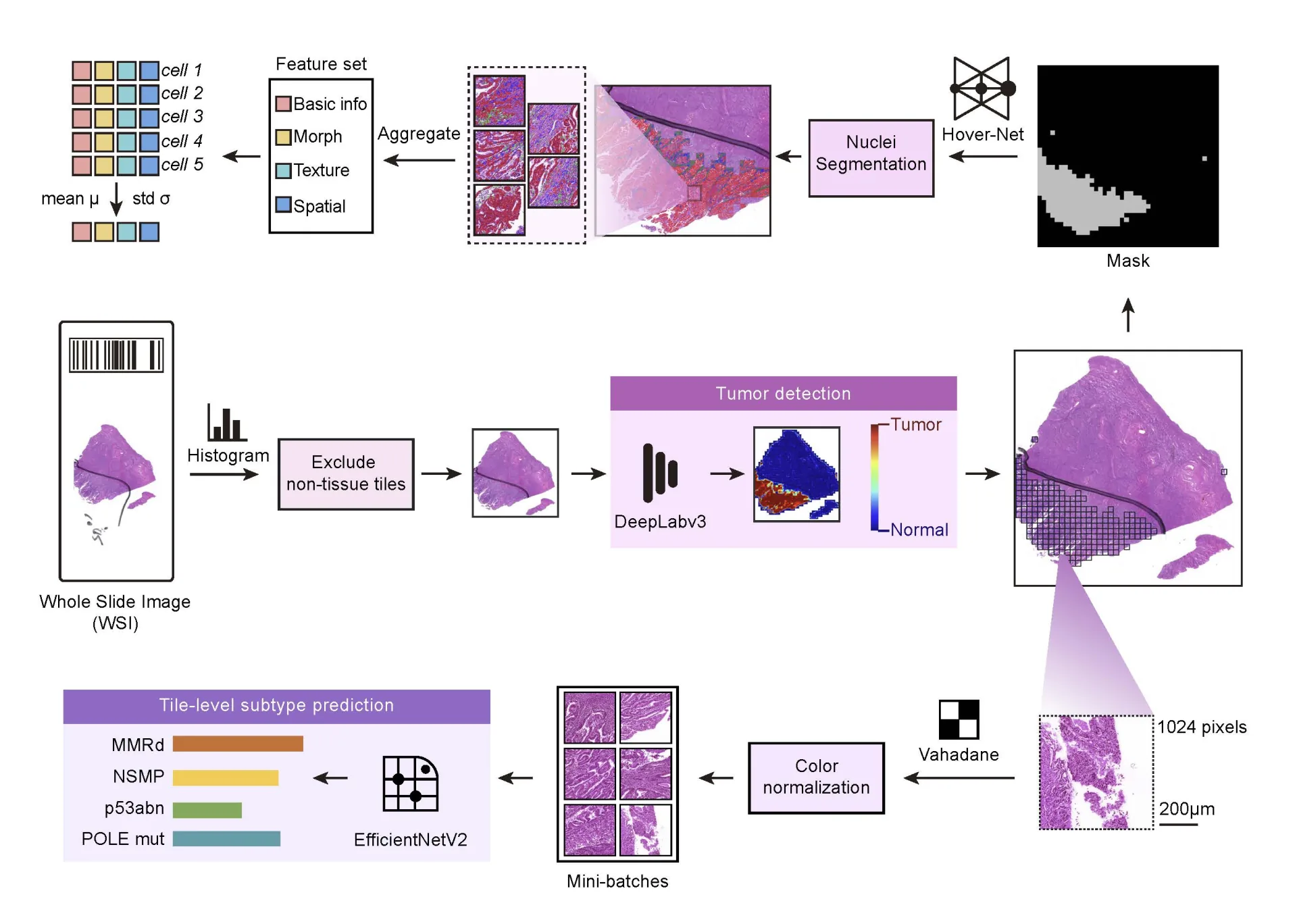
Figure. Schematic diagram of the model process proposed in this article
Reference method and original text link of this article:
Guo, Q., Cui, H., Zhang, Y. et al. An interpretable deep learning model for predicting endometrial cancer molecular subtypes from H&E-stained slides. npj Precis. Onc. (2026). https://doi.org/10.1038/s41698-026-01280-w
Translated from the original Chinese source.