The intelligent diagnosis model based on biopsy pathology developed by Dr. Chengfei Cai of IMIC Lab can significantly improve the clinical diagnosis efficiency of inflammatory bowel disease.
- IMIC Lab
- News , Comprehensive news
- July 14, 2025
The paper “IBDAIM: Artificial intelligence for analyzing intestinal biopsies pathological images for assisted integrated diagnosis of inflammatory bowel disease” by Dr. Cai Chengfei of Jiangsu University Key Laboratory of Intelligent Medical Image Computing (IMIC) was accepted by the International Journal of Medical Informatics. This result is the latest research result of cross-medical collaboration between the IMIC Lab (Professor Xu Jun’s team) and the Pathology Department of Drum Tower Hospital Affiliated to Nanjing University School of Medicine (Director Sun Qi’s team). It provides a new AI solution for the auxiliary diagnosis of inflammatory bowel disease. Current research in the field of computational pathology has made significant progress in the field of tumor pathology, but there are still obvious deficiencies in research on non-neoplastic lesions (especially inflammatory lesions). In particular, the identification and detection of intestinal inflammatory lesions face many challenges.
Research background
Inflammatory Bowel Disease (IBD) is a chronic gastrointestinal disease whose pathogenesis remains unclear. Its main subtypes include Crohn’s Disease (CD) and ulcerative colitis (UC). IBD can occur at any age, but it mainly affects young adults. Its clinical manifestations vary greatly, there is a lack of specific treatments, the disease course is protracted, and the incidence of complications is high, which brings a heavy economic burden to patients, their families and society. In recent years, the incidence of IBD has been significantly increasing, which has become a problem that cannot be ignored in daily clinical work. Symptoms include abdominal pain, diarrhea, rectal bleeding, anorexia, and fatigue, which significantly affect patients’ quality of life. Although UC and CD share many symptoms, differential diagnosis has important clinical implications due to differences in treatment strategies, outcome prediction, comprehensive evaluation, and clinical care. The current clinical manifestations of IBD are complex and the misdiagnosis rate is high. Accurate diagnosis relies on a combination of clinical manifestations, endoscopy, histological diagnosis, laboratory examinations and imaging examinations. Therefore, accurate diagnosis of IBD is a prerequisite and basis for treatment, and auxiliary diagnosis of IBD can reduce the differences between diagnoses by different pathologists.
Histological examination is an important component in ensuring accurate diagnosis and appropriate treatment of many diseases. In routine practice, it involves visual assessment of key histological and cellular patterns in tissues, an important step in understanding various disease states. Pathological examination plays a crucial role in the diagnosis, treatment, and follow-up monitoring of IBD patients. Pathological examination has also been widely used in medical institutions around the world, but the diagnosis of IBD lacks qualified pathologists. Pathologist resources are unevenly distributed, causing primary medical institutions to face certain difficulties in diagnosing IBD. The current diagnostic level of pathologists is uneven, which may lead to missed or misdiagnosed pathological examination results and delayed treatment. Detecting atypicals as lesions not only increases costs for patients but also increases the risk of disease progression. Therefore, in pathological diagnosis, how to reduce false positives and improve pathologists’ IBD pathological diagnosis is an urgent problem that needs to be solved. Perhaps the best way to solve the current problem is through highly sensitive artificial intelligence-assisted systems. Advanced artificial intelligence-assisted systems will improve diagnostic accuracy and efficiency, facilitate precise diagnosis and treatment, and improve patients’ quality of life.
research methods
Aiming at the problem of inflammatory type diagnosis in intestinal biopsy pathological images, this paper proposes an auxiliary diagnosis paradigm for effective fusion of multi-stage feature information, aiming to distinguish abnormal biopsy and inflammatory biopsy types, assist histopathological examination, and improve the diagnostic level of pathologists. The method proposed in this article was verified on data sets from two centers, proving that the inflammatory bowel disease biopsy pathological image diagnostic model (IBDAIM) proposed in this article can be used for the screening of normal intestinal biopsies and the diagnosis of major IBD types. By using internal data set cohorts and cross-validation methods to train the model, and then using internal and external validation cohorts to verify the diagnostic accuracy of the model, scientific evaluation methods are used to fairly evaluate the model’s improvement in the diagnosis results of pathologists.
The diagnostic model constructed in this article is a weakly supervised learning model that assists pathologists in diagnosis. The main process is divided into steps: image block level prediction and panoramic slice (WSI) level prediction. This paper first segments biopsy WSI into small image patches for prediction. A multiple-instance learning (MIL) algorithm is then utilized to fuse image patch features to obtain WSI-level predictions. Specifically: there are mainly the following four parts. (1) Diagnostic model training at the image patch level: This paper proposes that the comprehensive diagnostic model of inflammatory bowel disease inflammation types is a three-classification problem, in which each WSI is divided into one of the types such as normal intestinal tissue, UC, or CD. The model proposed in this paper is used to train three-class classification of image patches in WSI by training a deep convolutional neural network in a training queue. This article also similarly constructed a two-classification model, namely a diagnostic model for normal large intestine tissue and IBD, and a diagnostic model for UC and CD. This will facilitate the evaluation of the difference between the performance of this paper’s diagnostic model and the pathologist and quantify the gain of the model in assisting the pathologist’s diagnosis. ; (2) WSI fusion based on multi-instance learning: The image block level prediction model performs label prediction and probability calculation for all image blocks. This paper uses the MIL method to aggregate the probabilities of these image patches to train a classifier to generate WSI-level predictions. In order to collect the likelihood probability of image patches, this paper adopts two different machine learning methods, namely image patch likelihood histogram (PLH) and bag of words (Bag of Words, BoW) strategy. By using these two independent pipelines, we effectively fuse the scattered patch-level predictions to generate WSI-level features that are used in subsequent analysis operations. (3) Feature selection and classifier: Due to the high dimensionality of the acquired features, direct training of the classifier will lead to increased computational complexity, the risk of overfitting, poor model generalization, and reduced model interpretability. This article uses the Pearson correlation coefficient to evaluate the feature correlation coefficient, and then selects features with higher correlation coefficients for cluster analysis. After filtering out the relevant features, use the LASSO regression model for feature selection. The optimization goal is to minimize the sum of the loss function and the norm penalty term. In the final prediction, the machine learning algorithm model used in this article realizes differential diagnosis. At the same time, this article uses multiple classification algorithms for comparison. (4) The result gain of model-assisted pathologist diagnosis: For the internal test set and the external test set, the pathologist conducts two rounds of diagnosis to quantify the clinical benefits that the pathologist actually obtains with the help of the IBDAIM model. Five pathologists with an average of 7 years of experience in intestinal pathology diagnosis (2-15 years) participated in this study. For the internal test set and the external test set, pathologists are randomly presented when making diagnoses.
experimental design
This study collected 543 tissue samples from 2014 to 2023 from the Affiliated Drum Tower Hospital of Nanjing University School of Medicine (NDTH). Each sample was made into multiple serial sections, placed on the same slide, and stained with H&E. Due to the high similarity between consecutive slices, this study uses consecutive samples as WSI to prevent data overlap when randomly generating the model set and the internal test set. This study used biopsy data from NDTH as the model training set. Three WSIs were excluded due to faded H&E staining and unclear scanning. The training data set includes normal tissue (184 WSIs), CD (182 WSIs), and UC (174 WSIs). In the classification task studied in this article, the data set is randomly divided into a model set and an internal test set with a ratio of 8:2. In addition, H&E stained biopsy data of tissue samples from Zhujiang Hospital of Southern Medical University were collected as an external validation set (ZJH), and a total of 305 WSIs were collected. Among them, there were 90 WSIs for normal extensive intestinal tissue, 113 WSIs for CD, and 102 WSIs for UC. All WSIs were digitized using an Aperio GT450 scanner at a resolution of 263 nm/pixel. Figure 1 is an overview of the research framework of this article.
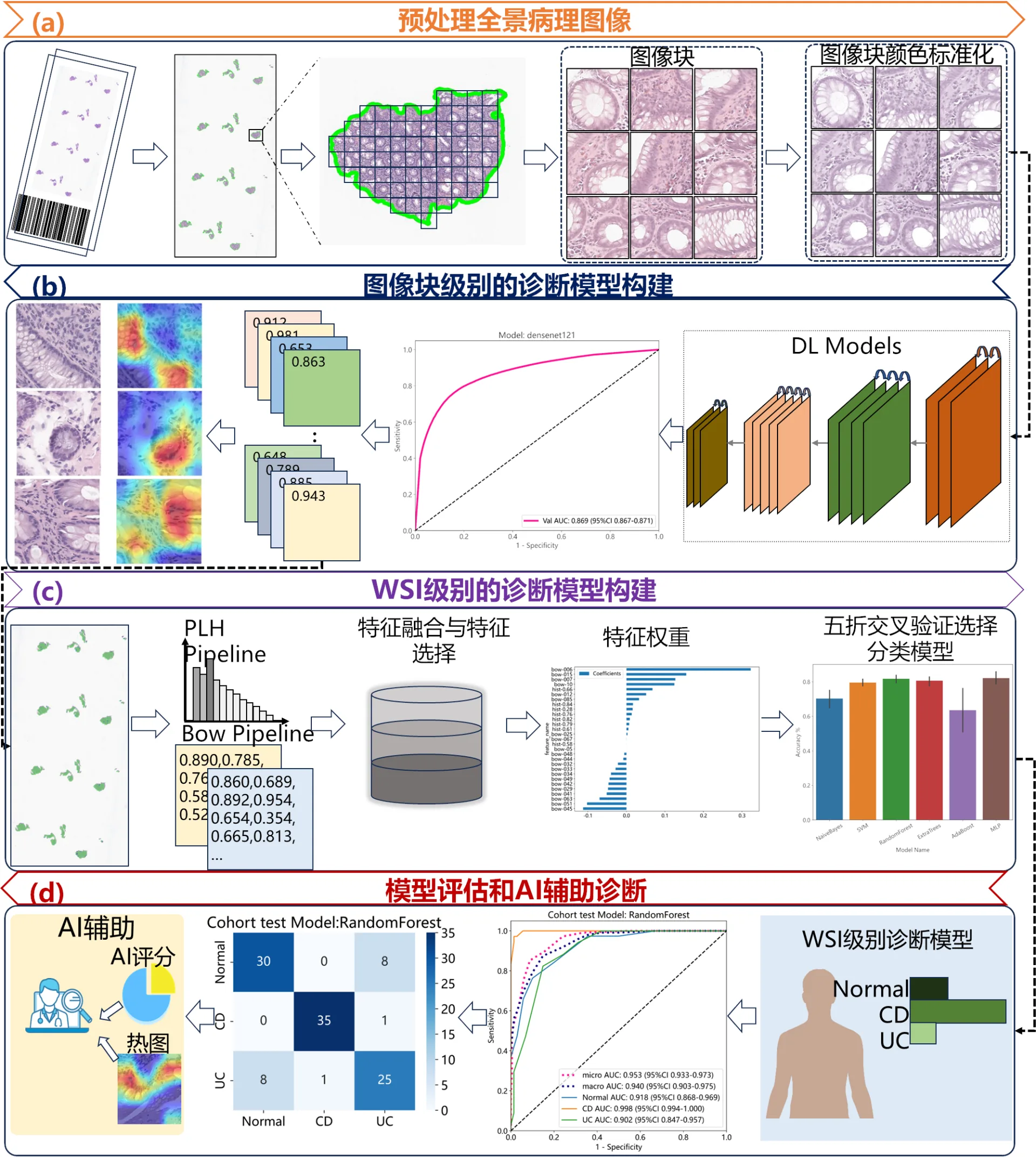
Figure 1: This article’s process framework for building an AI model to assist IBD diagnosis. (a) is to segment the WSI into image patches and perform color normalization processing; (b) is to build a diagnostic model at the image patch level; (c) first, use two sets of features to represent each image patch aggregation, and secondly, aggregate the image patch prediction results and represent them as features of the WSI. Then use feature selection method to select effective features, and finally use five-fold cross-validation to build a WSI-level diagnostic model. (d) Test the constructed diagnostic model on the test set and assist the pathologist in diagnosis, and at the same time evaluate the gain of the model in the pathologist’s auxiliary diagnosis.
Experimental results
In building a binary classification diagnostic model at the WSI level, SVM is used to build a classification model. This study achieved excellent results by building a model using aggregated features of BoW and PLH. This study generates confusion matrices for training and test datasets for binary classification tasks to intuitively explain model classification performance. Figure 2 shows the results of this study on the internal test set and the external test set in the two tasks of two classifications. In the task of distinguishing normal from inflammation, the model performed better on the test set. In the task of distinguishing UC and CD, CD is easily misclassified as UC, while UC is usually judged as UC.
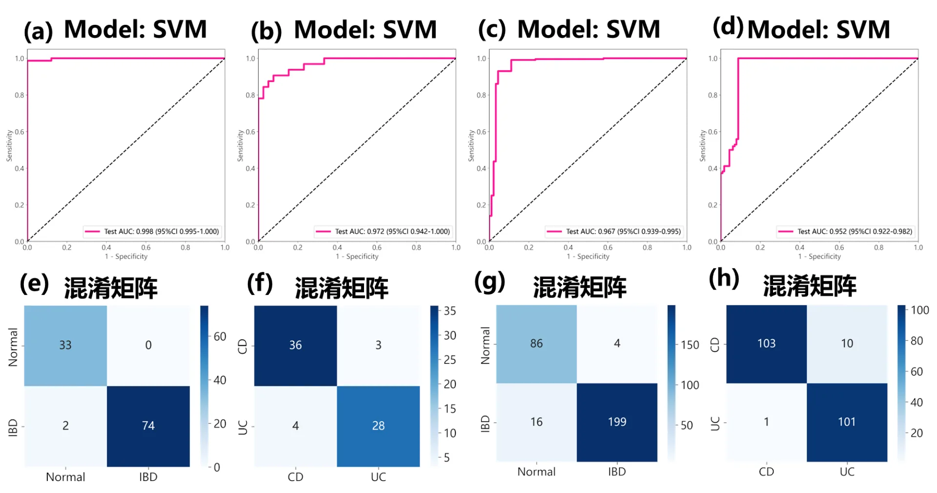
Figure 2: AUROC and confusion matrix for the binary classification task in the inner and outer test sets. AUROC and confusion matrix of Normal vs. IBD task in (a), (c), (e), (g), where (a) and (e) are the results of the internal test set and (c) and (g) are the results of the external test set. (b), (d), (f), (h) are AUROCs in the CD vs. UC task
and the results of the confusion matrix, where (b) and (f) are the results of the internal test set, (d) and (h) are the results of the external test set.
In the evaluation of multi-classification model diagnostic efficiency, this study tested 6 classification models, among which the random forest classifier showed the most accurate classification results. Figure 3 shows the AUROC and confusion matrix of the random forest classifier on the training set, inner test set, and outer test set. Judging from the results of the test set, the model has relatively reliable performance for normal, CD and UC diagnostic models.
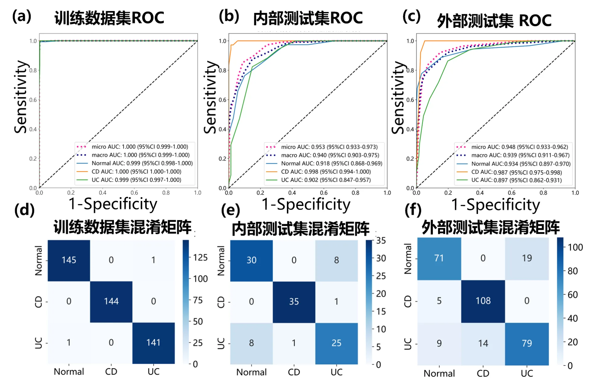
Figure 3: Shows three types of AUROCs and confusion matrices for the training set, inner test set, and outer test set. (a) and (d) are the AUROC and confusion matrix (b) of the model in the training set
and (e) are the AUROC and confusion matrix of the model in the internal test set; (c) and (f) are the AUROC and confusion matrix of the model in the external test set.
In this study, five pathologists were selected to conduct two rounds of diagnosis on all test sets, namely diagnosis alone and diagnosis with the assistance of the IBDAIM model. The diagnosis of IBD is very challenging, and the diagnostic accuracy of junior pathologists is low. In actual clinical practice, experienced pathologists can diagnose IBD with a high accuracy with the support of pathological slides and sufficient clinical information. The diagnosis results of each pathologist in different cohorts in the first round of diagnosis are shown in the human row of Figure 4 . Pathologists are more accurate in diagnosing the presence or absence of inflammation. In the task of distinguishing UC and CD, the diagnostic accuracy of junior pathologists was much lower than the accuracy of the model, and the diagnostic accuracy of experienced pathologists without the support of clinical information was also slightly lower than the accuracy of the model. The AI-assist row of Figure 4 shows that in the second round of diagnosis with the assistance of the IBDAIM model, the diagnostic accuracy of the young pathologist is close to that of the experienced pathologist, while the diagnostic accuracy of the experienced pathologist reaches or even exceeds the model accuracy. Additionally, Figure 5 shows the average diagnostic performance improvement of pathologists with AI assistance.
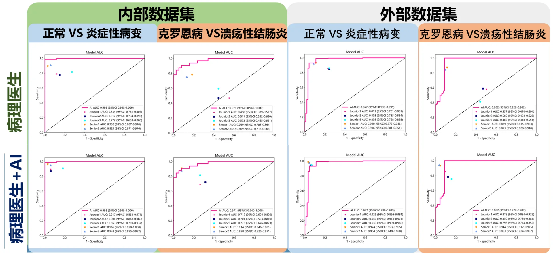
Figure 4: IBDAIM assists pathologists in improving the efficiency of intestinal biopsy tissue diagnosis.
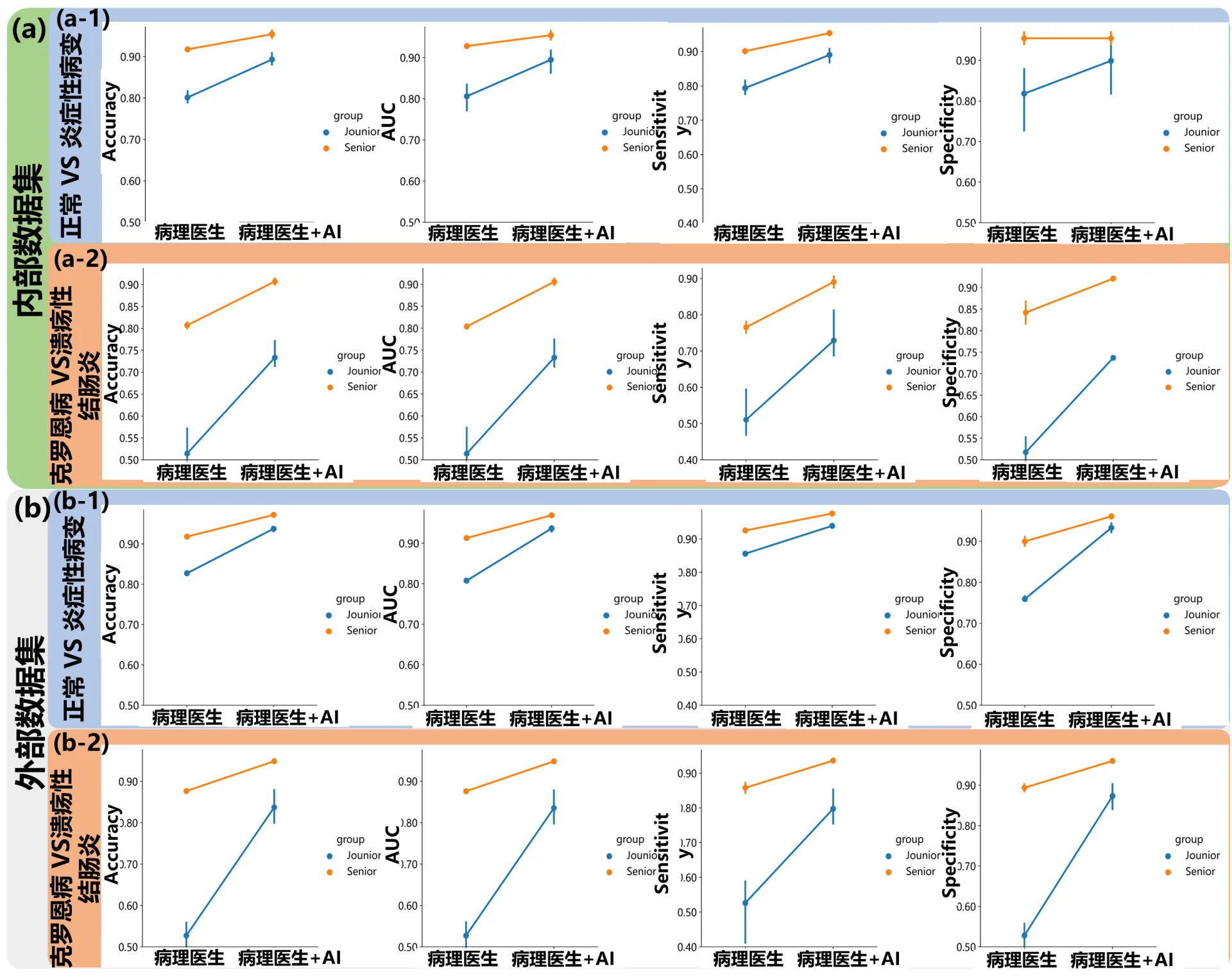
Figure 5: Average diagnostic performance improvement by pathologists using IBDAIM-assisted diagnosis on internal and external test sets. (a) Shows the performance of pathologists with different experiences on the IBDAIM-assisted in-house test set.
Average diagnostic performance improvement under assisted diagnosis. (b) Shows the average diagnostic performance improvement of pathologists with different experience under IBDAIM-assisted diagnosis on the external test set.
Conclusion
The IBDAIM model developed in this study shows excellent performance in the diagnosis of normal, CD and UC, giving it important clinical application potential in assisting pathologists in the diagnosis of IBD. Currently, pathologists have low efficiency and accuracy in diagnosing various types of IBD, and there is a lack of experienced pathologists. This article provides an in-depth discussion of the role of the IBDAIM model in improving the diagnostic efficiency of pathologists. Pathologists trained with 2-15 years of pathology diagnosis experience had higher accuracy in distinguishing normal from IBD, slightly lower than the model’s performance. However, there was a significant decrease in the diagnosis of CD and UC. This phenomenon may stem from the fact that in clinical practice, accurate diagnosis of CD and UC often relies on comprehensive analysis of multi-modal examination information, while diagnosis based solely on H&E stained pathological slides, especially for pathologists with limited experience, cannot guarantee accuracy. After AI-assisted diagnosis, the pathologist can obtain the probability value predicted by the model and the heat map of the model’s area of interest from the model prediction results, thereby assisting the pathologist in observing subtle diagnostic characteristics that are difficult to find. Experimental results show that the IBDAIM model has the potential to become a diagnostic tool for IBD biopsy pathology images. The model can not only assist pathologists to improve diagnostic efficiency, but also provide guidance for young pathologists and effectively reduce the workload of senior pathologists. By excluding non-abnormal samples, the model optimizes the diagnostic process of inflammatory bowel disease, thereby improving the overall diagnostic efficiency.
The citation information for this article is as follows:
Chengfei Cai, Qianyun Shi, Mingxin Liu, Jun Li, Yangshu Zhou, Andi Xu, Dan Zhang, Yiping Jiao, Yao Liu, Xiaobin Cui, Jun Chen, Jun Xu, Qi Sun, IBDAIM: Artificial Intelligence for Analyzing Intestinal Biopsies Pathological Images for Assisted Integrated Diagnostic of Inflammatory Bowel Disease, International Journal of Medical Informatics , vol. 203, 2025, 106024.
Translated from the original Chinese source.