Digital Pathology + Artificial Intelligence: 400 Million Cell Atlas Analyzes Breast Cancer Ecotypes
- IMIC Lab
- News , Comprehensive news
- October 26, 2023
From a microscopic perspective, a towering tree is not just the tree itself, but a huge ecosystem composed of many different organisms and their complex interactions. Similarly, breast cancer is not only composed of tumor cells, but also a huge ecosystem composed of many different cells such as inflammatory cells and stromal cells and their complex interactions. The biological behavior, prognosis and efficacy of breast cancer depend not only on the characteristics of tumor cells, but also on other cellular components and their interactions with tumor cells. Analyzing the composition of the breast cancer ecosystem and the relationship between different types of cells is of great significance for understanding the biological behavior of breast cancer and improving the level of accurate diagnosis and treatment of breast cancer. Since cells are the basic unit of life, they are also the basis for the occurrence and development of diseases. Traditional pathology can only perform descriptive analysis on a small number of cells using hematoxylin and eosin (H&E) stained sections and optical microscopy; as digital pathology is adopted on a large scale in clinical practice, we can use advanced data and knowledge-driven machine learning technology and digital image processing technology to carry out multi-omics and massive data on hundreds of millions of cells and their tissue structures in high-resolution digital pathology sections. Quantitative analysis can reveal the trends and patterns of disease occurrence and development, thereby helping us better understand the disease and assist doctors in formulating optimal treatment strategies.
On October 25, 2023, the paper “Single cell morphology and topological maps reveal the ecosystem diversity of breast cancer”, a collaboration between Professor Xu Jun’s team at the Smart Medical Research Institute and Professor Shao Zhimin’s team at Fudan University Cancer Hospital, was published online in the “Nature Communications” magazine of the British “Nature”. Chen Depin, a master’s student at the Smart Medical Research Institute, and Zhao Shen, Fu Tong, and Yang Jing from Fudan University Cancer Hospital became the co-first authors of this article. Professor Shao Zhimin and Professor Jiang Yizhou from the Breast Surgery Department of Fudan University Cancer Hospital, Professor Yang Wentao from the Department of Pathology, and Professor Xu Jun from the Smart Medical Research Institute are the co-corresponding authors of this article. With the support of major research projects of the National Natural Science Foundation of China, multidisciplinary teams such as the Department of Breast Surgery and Pathology of Fudan University and the Artificial Intelligence of the Smart Medical Research Institute collaborated in depth, integrating multidisciplinary knowledge such as machine learning, mathematical graph theory, and digital image processing to create the first single-cell tumor morphology and topology analysis algorithm (sc-MTOP). A single-cell map of breast cancer containing 410 million cells was drawn, comprehensively depicting the diversity of the breast cancer ecosystem, and providing a basis for digital pathology and artificial intelligence. Empowering precise diagnosis and treatment of breast cancer provides a new perspective.
First, the researchers applied sc-MTOP to 637 cases of breast cancer (405 cases of hormone receptor positive and HER2 negative, 85 cases of hormone receptor positive and HER2 positive, 66 cases of hormone receptor negative and HER2 positive, and 81 cases of triple negative) in the Tumor Hospital of Fudan University for theorem analysis. The nuclear morphology and cell spatial distribution relationship characteristics of single cells were extracted from approximately 410 million cells, allowing for quantitative analysis of the breast tumor ecosystem.
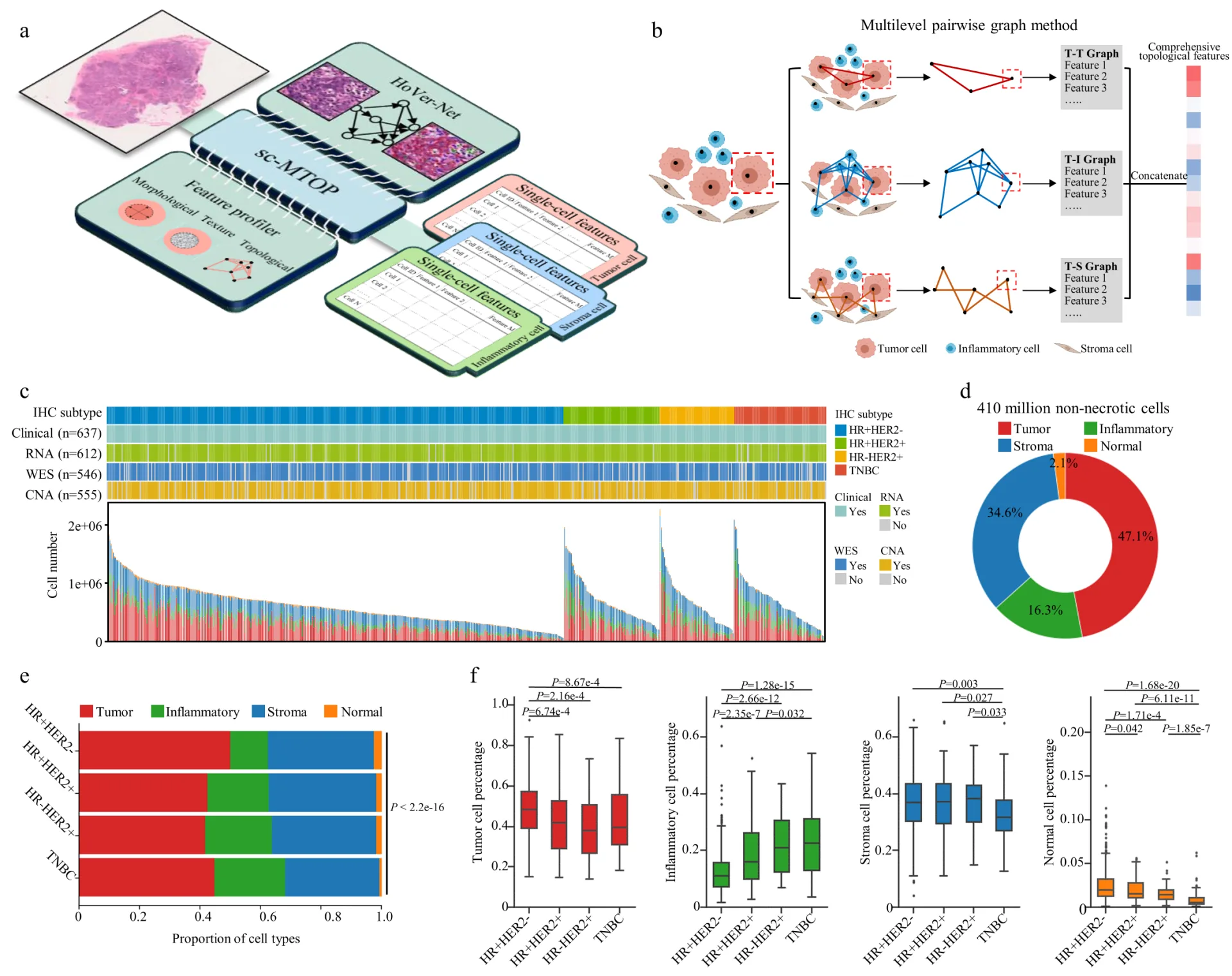
Figure 1: sc-MTOP analysis and its generated data set
Subsequently, the study constructed a single cell atlas of 410 million cells, including 190 million tumor cells (47.1%), 66.85 million inflammatory cells (16.3%), 140 million stromal cells (34.6%), and 8.6 million normal breast cells (2.1%), and analyzed the phenotypic diversity of tumor cells, inflammatory cells, and stromal cells respectively.
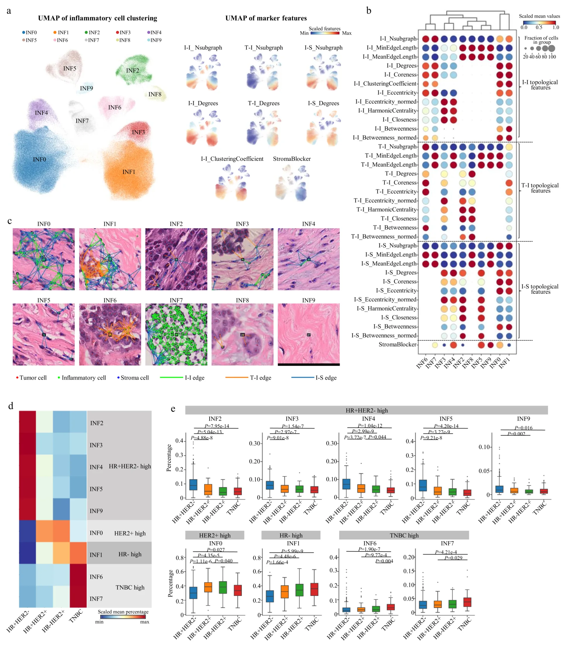
Figure 2: Single-cell atlas of inflammatory cells
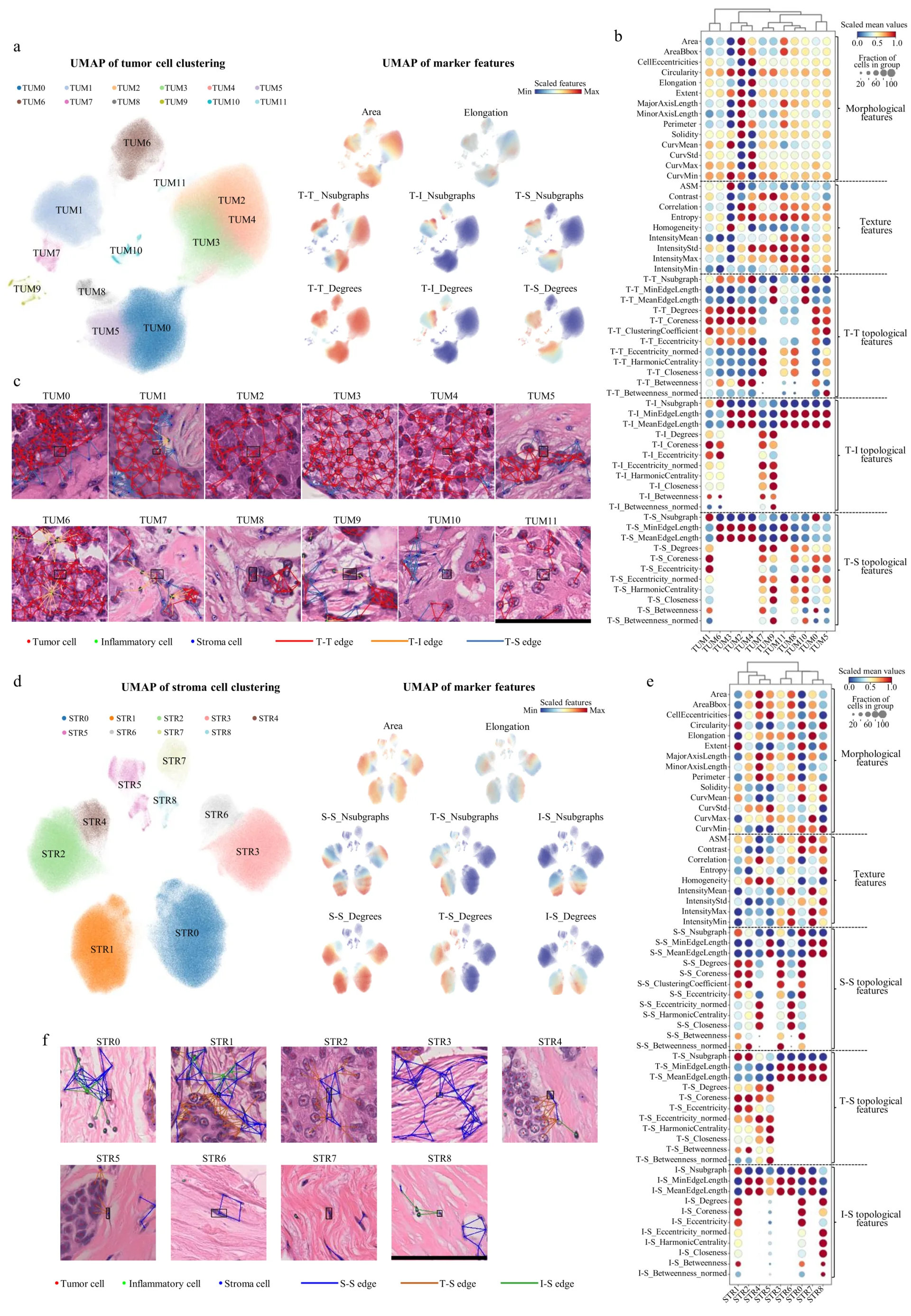
Figure 3: Single cell atlas of tumor cells and stromal cells
Finally, this study analyzed microecological patterns representing local multicellular structures and recurrence-free survival through spatial distribution, revealing four breast cancer ecotypes with different molecular characteristics associated with patients’ recurrence-free survival.
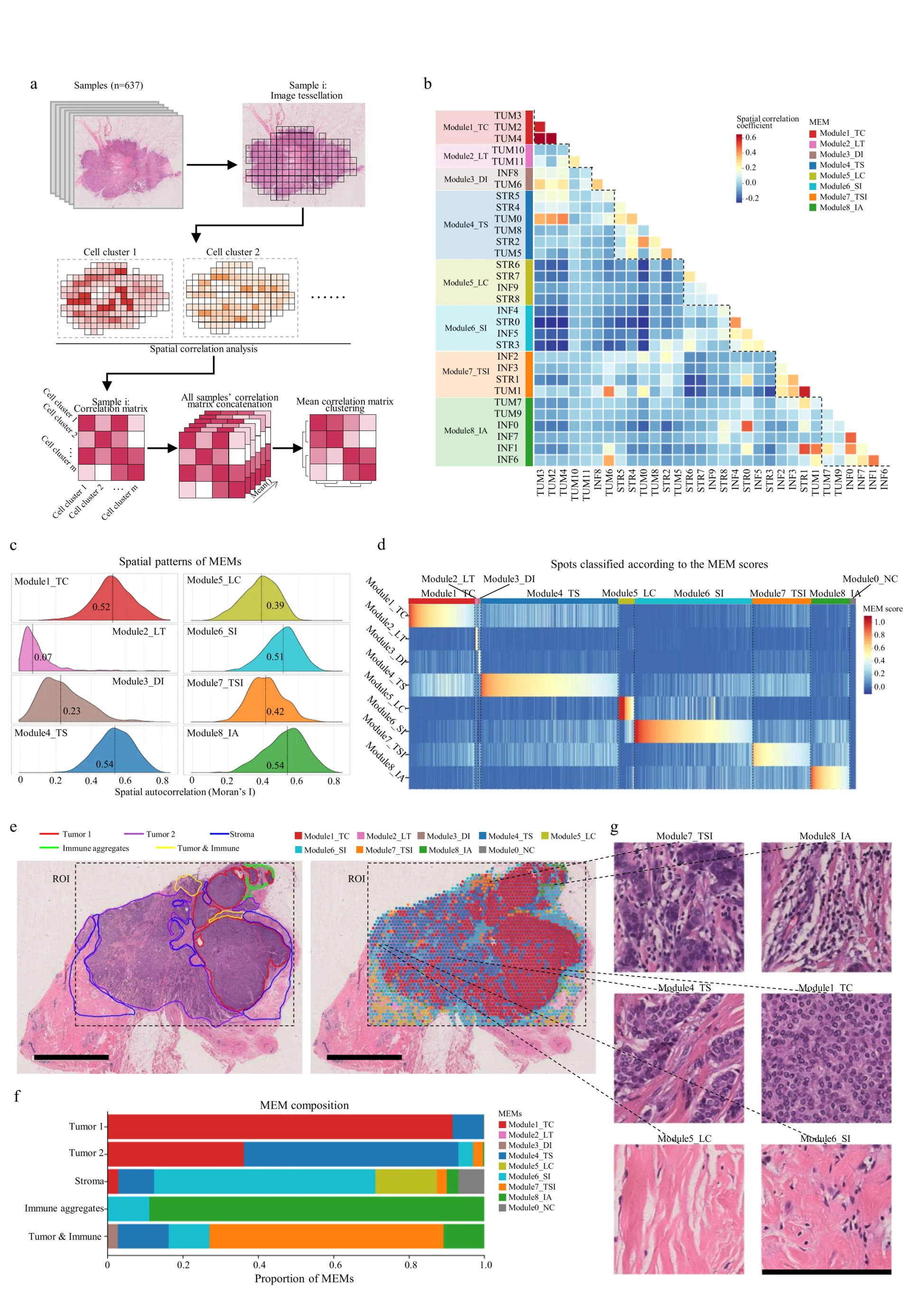
Figure 4: Single-cell atlas of tumor cells and stromal cells
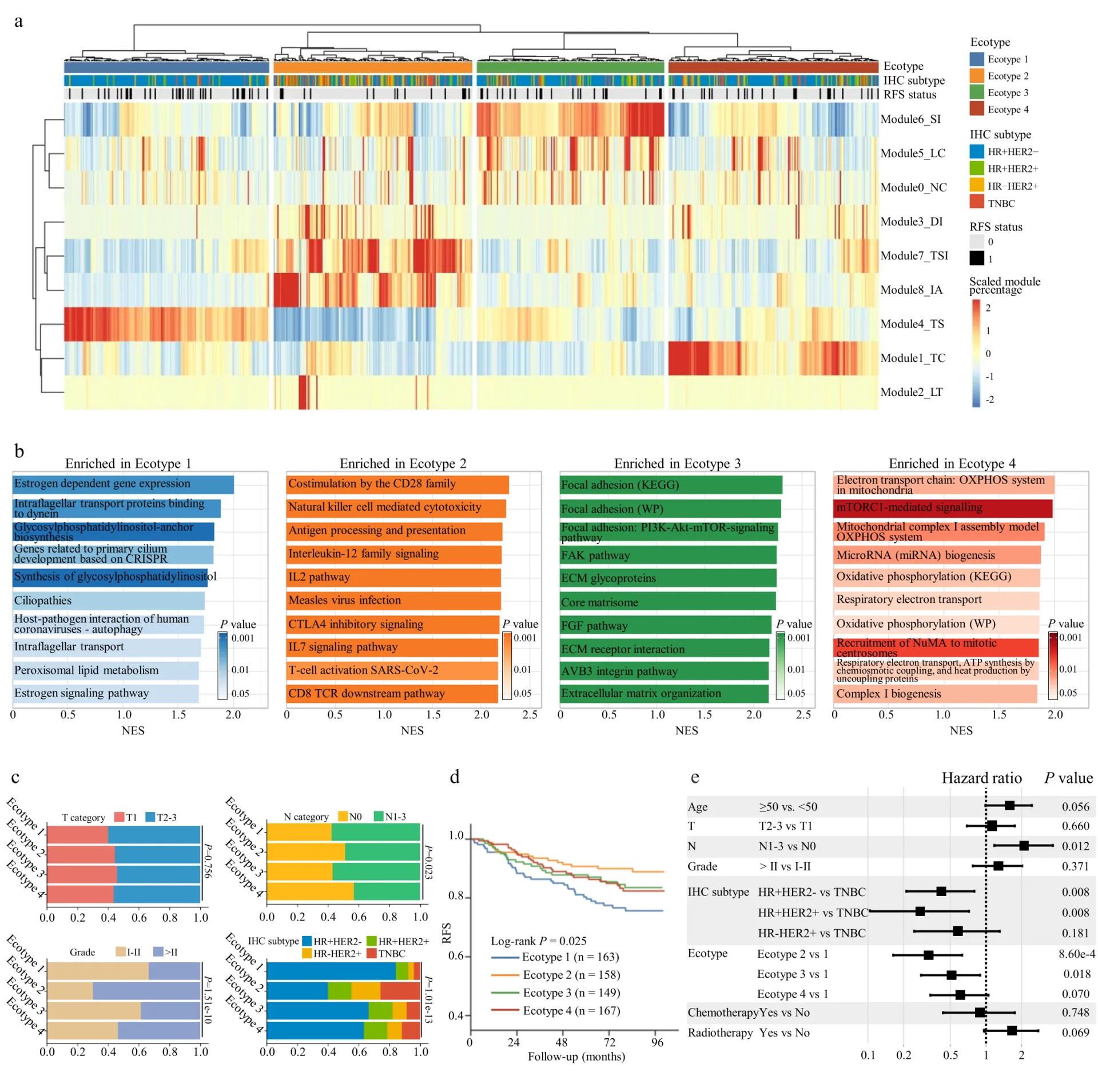
Figure 5: Breast cancer ecotypes with different microbial patterns are associated with molecular characteristics and patient recurrence-free survival.
Further analysis of multi-omics data revealed clinically significant ecosystem characteristics: Triple-negative breast cancer has a large number of local accumulation of inflammatory cells, which can indicate immune activation in the tumor microenvironment, which is beneficial to the effect of immunotherapy; hormone receptor-positive breast cancer has tumor cell nuclear morphological heterogeneity, which can indicate cell cycle pathway activation and the efficacy of CDK inhibitors.
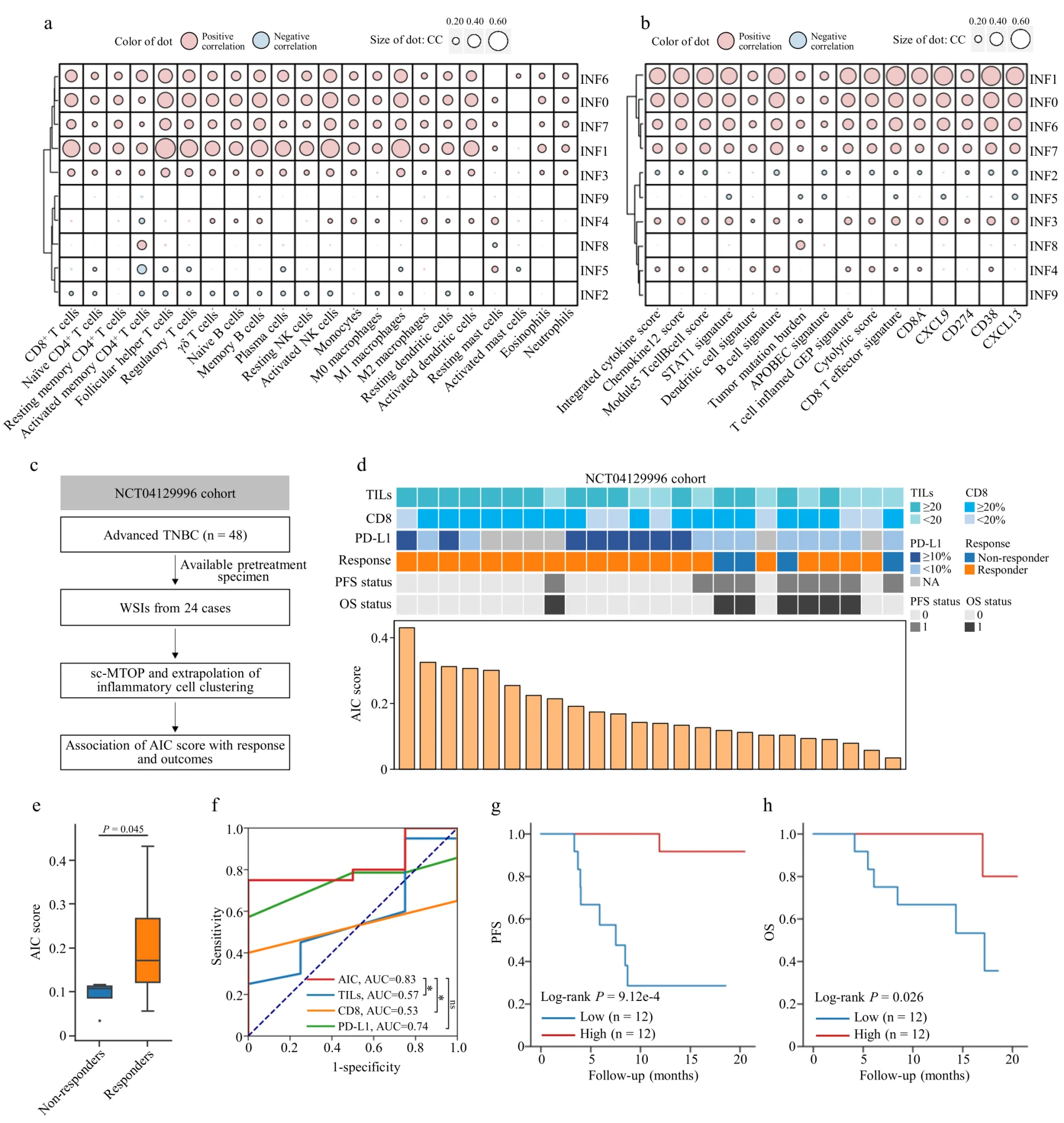
Figure 6: The local accumulation and abundance of inflammatory cells in triple-negative breast cancer can indicate the effect of immunotherapy
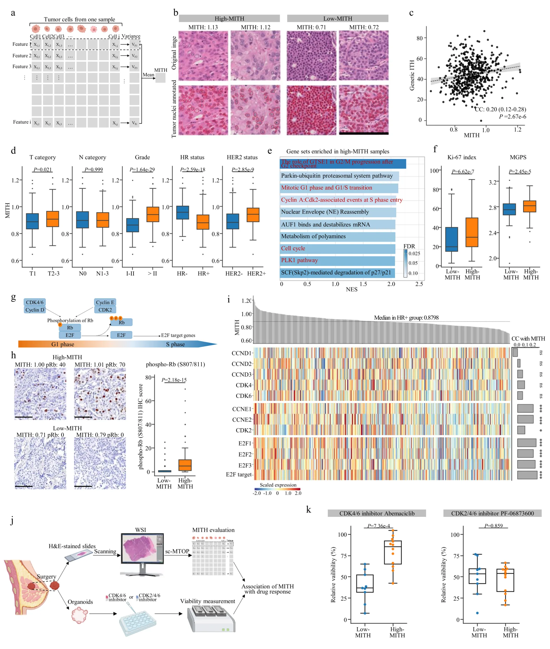
Figure 7: Nuclear morphological heterogeneity in hormone receptor-positive breast cancer tumors can indicate cell cycle pathway activity and CDK inhibitor efficacy.
Therefore, the results of this study show that the sc-MTOP algorithm can analyze the tumor ecosystem from the single cell level on digital pathology section images. Digital pathology combined with artificial intelligence has become a unique research direction and important transformation tool under the breast cancer accurate classification system of Fudan University Cancer Hospital.
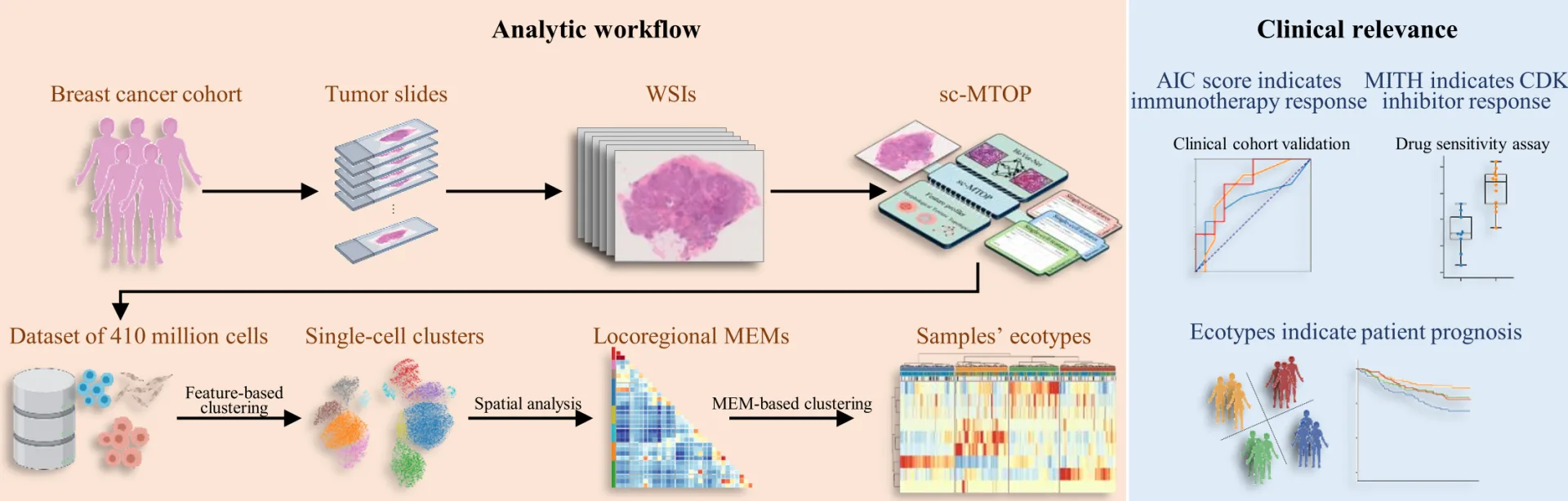
Figure 8: Research summary
The citation information of the paper is as follows:
Zhao, S., Chen, DP., Fu, T. et al. Single-cell morphological and topological atlas reveals the ecosystem diversity of human breast cancer. Nat Commun 14, 6796 (2023). https://doi.org/10.1038/s41467-023-42504-y
Translated from the original Chinese source.