Congratulations to Lu Haoda for successfully passing the defense of his doctoral thesis
- IMIC Lab
- News , Comprehensive news
- January 12, 2023
On the morning of January 10, 2023, Lu Haoda from the Smart Medical Research Institute successfully passed the defense of his doctoral thesis entitled “Automatic staging of mouse seminiferous ducts based on histomorphological analysis”. Members of the defense committee include Professor Chen Yang from the School of Computer Science and Engineering of Southeast University, Professor from the Singapore Science and Technology Agency (A*Star) ) Professor Yu Weimiao from the Institute of Bioinformatics, Professor Fan Xiangshan from the Pathology Department of Nanjing University Drum Tower Hospital, Professor Xu Yujun from the Reproductive Science Research Center of Northwestern University School of Medicine and the State Key Laboratory of Reproductive Medicine at Nanjing Medical University, Professor Li Chen from the School of Medicine and Bioinformatics Engineering at Northeastern University, Professor Fu Zhangjie and Professor Yuan Xiaotong from the School of Computer Science at Nanjing University of Information Science & Technology, and Associate Professor Wang Xiangxue from the School of Artificial Intelligence (School of Future Technology) at Nanjing University of Information Science & Technology. Professor Chen Yang served as the defense chair. Teacher Jiao Yiping is the defense secretary. The backgrounds of the defense committee members span across multiple disciplines such as clinical medicine, reproductive medicine, biomedicine, computer science, and artificial intelligence. The members come from three countries: China, the United States, and Singapore. The completion of Lu Haoda’s doctoral thesis defense marks that the Smart Medical Research Institute’s research in the field of reproductive medicine has entered a new stage.
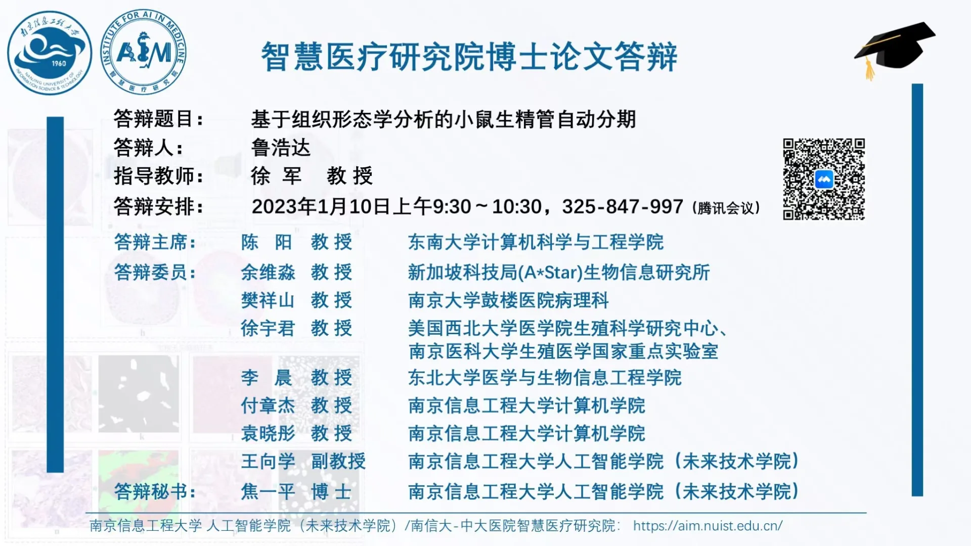
Defense poster
Lu Haoda’s doctoral thesis established for the first time a sophisticated intelligent staging system based on histological morphology based on H&E stained mouse seminiferous duct images. The main work includes:
As the basis of the fine staging system, strategies such as edge overlapping sliding windows, multi-scale learning, and multi-task learning are proposed to address issues such as seminiferous duct segmentation, initial staging semantic segmentation, and multi-type germ cell segmentation, which effectively improve the segmentation performance, robustness, and generalization of each task;
Aiming at the problem of fine staging within each group of early, middle and late seminiferous ducts, we use prior knowledge of histopathological morphology to extract rich quantitative features such as cell morphology, spatial topology, cell direction and texture, and combine feature selection algorithms and machine learning models to achieve automatic staging. The staging accuracy has reached the level of a pathologist with five years of experience.
In specific fine staging problems, DtG filters, pathology-independent and pathology-related self-supervised auxiliary tasks, texture feature visualization, Grad-CAM and other technologies are introduced to effectively improve the accuracy and interpretability of the staging system.
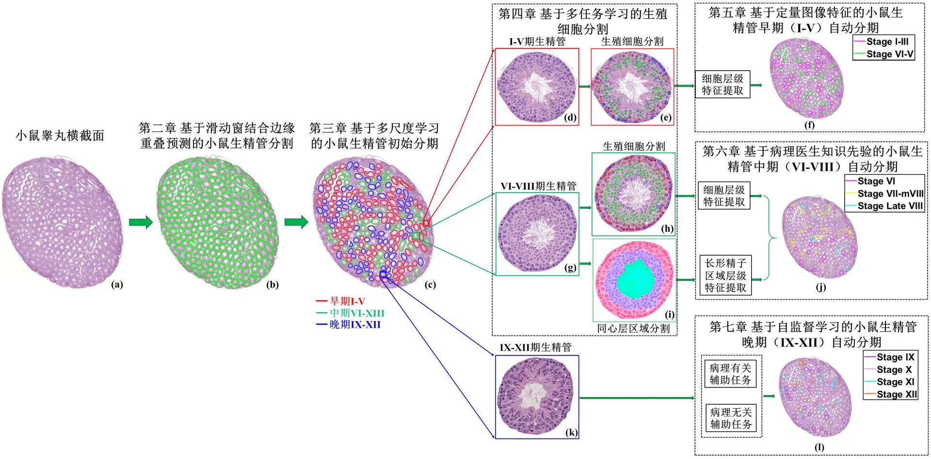
Chapter structure of Lu Haoda’s doctoral thesis
This fine staging system can be used in the field of assisted reproductive medicine to conduct quantitative research on male infertility problems caused by defects in seminiferous duct development, and also provides new solutions for other computational pathology studies. In the future, further research will be conducted on the seminiferous ducts of genetically defective mice to explore the complex mechanisms behind spermatogenic development defects.
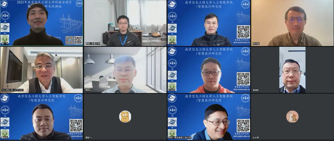
Group photo of defense experts
The institute’s work on seminiferous duct section analysis originated in early 2017. Professor Xu Jun from the Smart Medical Research Institute and Professor Xu Yujun from the State Key Laboratory of Reproductive Medicine at Nanjing Medical University had an accidental exchange at an academic conference, which led to this cooperation. This series of work is the first to establish an intelligent seminiferous duct automatic staging system for conventional H&E sections of mouse testicles based on quantitative histomorphological analysis. At present, most work in the field of computational pathology focuses on tumor pathology. This work is oriented to non-tumor pathology and is very challenging. Many technical routes are attempted for the first time. It is also a pioneering work in pathological image analysis of seminiferous duct tissue. Compared with current analysis methods based on fluorescence imaging, this work is oriented to conventional H&E sections and has better practicability and generalizability, but its technical difficulty increases sharply.
After more than 5 years of hard research and perseverance, Lu Haoda overcame many difficulties and successfully established an automatic analysis system for mouse seminiferous ducts based on H&E. This series of work has attracted the attention of Rex A Hess, a famous expert in male reproductive pathology at UIUC in the United States, and the team of Professor Don Conrad, a famous scholar in the field of male infertility genetic analysis at Oregon Health and Science University. After the paper was published, it received MIT-Harvad Multidisciplinary Institute of Integration, MIT Department of Electrical Engineering and Computer Science, Department of Data Science, Department of Pathology and Department of Biostatistics of Harvard University, Department of Psychiatry at Massachusetts General Hospital, Department of Stem Cell and Regenerative Biology at Harvard University, Department of Genetics at the National Primate Research Center (one of the seven federally funded national research centers in the United States) at Oregon Health and Science University, Department of Obstetrics and Gynecology, Department of Pathology, Department of Psychology, Semmelweis University in Hungary, and other multidisciplinary experts.
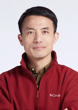
State Key Laboratory of Reproductive Medicine, Nanjing Medical University & Professor Xu Yujun, Northwestern University School of Medicine
The completion of Lu Haoda’s doctoral thesis received help and support from many scholars, first of all, the long-term support of Professor Xu Yujun and his team. Professor Xu Yujun is a Distinguished Professor of Jiangsu Province and a Distinguished Professor of Nanjing Medical University; an adjunct professor of the Department of Obstetrics and Gynecology and Psychiatry and Behavioral Sciences of Northwestern University; a leading talent in the scientific and technological innovation male reproductive medicine team of Jiangsu Province; a doctoral supervisor; and a well-known scholar in the field of male reproductive development. Professor Xu Yujun is committed to studying mammalian reproductive development and the regulatory mechanisms of stem cells, especially the highly conserved fundamental mechanisms and translational medical research on the role of reproductive-related diseases. Through comparative biology and genomic analysis of germ cell development in the animal kingdom, it was discovered that there are highly conserved core machines and components that regulate gametogenesis, including the DAZ gene family, the most common cause of male infertility. Currently, the functions of the DAZ family and other core members are being studied in depth through gene knockout and transgenic animal models (mouse, Drosophila), and their mechanisms of action in reproductive development and stem cell maintenance and differentiation are being analyzed.
Professor Rooij D.G. de Dirk, University of Utrecht & University of Amsterdam, Netherlands
This work also received the help and support of Professor Dirk from Utrecht University and the University of Amsterdam in the Netherlands. Professor Dirk is the most authoritative scholar in the world in the field of conventional H&E stained mouse testicular pathological staging. We all admire his seriousness and persistence in his work in his 70s. He is willing to use his lifelong accumulated experience and knowledge of seminiferous duct staging based on conventional H&E stained pathological sections for the training of the AI model. Although he has retired, he is still willing to learn to use pathological annotation software from scratch, and takes the trouble to carefully examine and mark the stages of a large number of seminiferous ducts one by one, providing extremely valuable training data for the construction of the model. Pathology is a “cold” field. Currently, there are very few experts who can accurately distinguish the development stages of seminiferous ducts I-XII based on conventional H&E stained pathological sections. Especially without the assistance of PAS staining, it is extremely difficult to accurately identify stages I-XII based only on conventional H&E sections. Professor Dirk is one of the few authoritative experts in this field in the world. Without his professional data annotation institute, it would be difficult to carry out a series of research in this field. For this reason, the annotated data used to train the model in the study contains Professor Dirk’s long-term accumulated experience and knowledge in analyzing the staging of seminiferous ducts.

Professor Anant Madabhushi, Emory University & Georgia Tech, USA
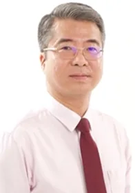
Professor Yu Weimiao, Agency for Science and Technology of Singapore (A*Star)
In addition, the smooth development of this work would not have been possible without the guidance and support at different stages from Professor Anant Madabhushi of Emory University & Georgia Tech University and Professor Yu Weimiao of the Agency for Science and Technology of Singapore (A*Star). The institute expresses its deep gratitude to all experts and scholars for their full support and looks forward to deeper and broader cooperation.
Lu Haoda graduated from Nanjing University of Information Science & Technology, majoring in automation and system science, with a bachelor’s degree in engineering and a master’s degree in science. In 2018, he was recommended to enter the School of Electronics and Information Engineering, Nanjing University of Information Science & Technology, to study for a doctorate in information and communication engineering. His supervisor is Professor Xu Jun, and his main research direction is histopathological image calculation. During this period, he was funded by the China Scholarship Council to conduct joint training at Nanyang Technological University in Singapore for one year. During Lu Haoda’s doctoral studies, he published two SCI Area 1 papers as the author or co-author, several SCI Area 2, 3, and Peking University core papers. He participated in many National Natural Science Foundation projects and won many scholarships and challenge awards. The defense committee and teachers and students of the graduate school extend warm congratulations to Lu Haoda!
Attached: The main papers published by the Smart Medical Research Institute in the field of reproductive medicine and their introductions
1 . Jun Xu, Haoda Lu, Haixin Li, Chaoyang Yan, Xiangxue Wang, Ming Zang, Rooij D.G. de Dirk, Anant Madabhushi, and Eugene Yujun Xu, “Computerized Spermatogenesis Staging (CSS) of Testis Sections for Mouse Sperm Development via Quantitative Histomorphological Analysis”, Medical Image Analysis , vol. 70, 101835, 2021.
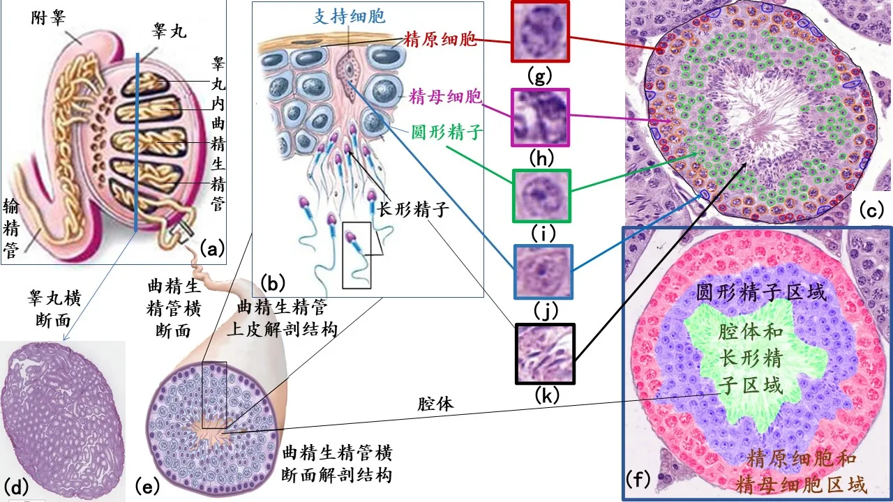
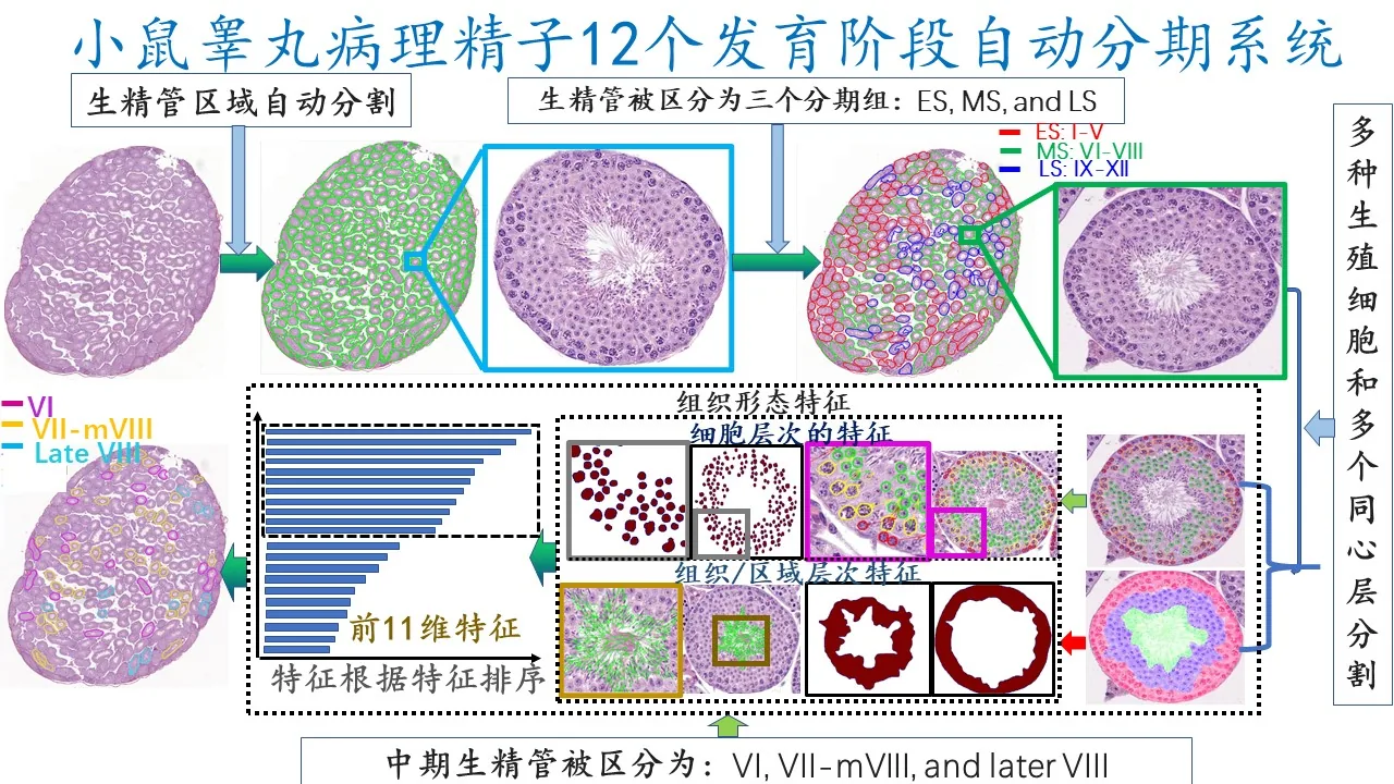
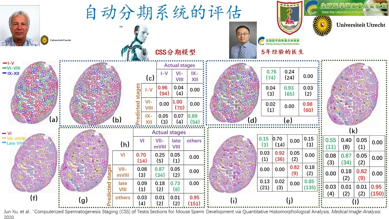
This work is the first to construct an automated staging system for 12 developmental stages of mouse testicular sperm based on histomorphological analysis of conventional H&E sections. Technically, this work is based on H&E sections, which are difficult to analyze without relying on PAS staining. This work developed a deep learning system for the segmentation of seminiferous ducts, accurate identification of multiple types of sperm cells and multiple pathological concentric layer regions for high-resolution panoramic slices. This research received full support from Professor Dirk in the data annotation and other stages. During the research process, in order to describe the unique pathological structure of the seminiferous tubules and the subtle characteristics at the cellular level, the paper tried and compared a large number of solutions, and finally succeeded. This work embodies the efforts of more than a dozen scholars and graduate students over many years. The researchers span the fields of biology, medicine, pathology, and engineering, as well as three countries/regions: China, Europe (Netherlands), and the United States. In-depth cross-disciplinary and cross-country collaboration contributed to this research, with all links playing a crucial role.
- Shi Liang, Haoda Lu, Min Zang, Xiangxue Wang, Yiping Jiao, Tingting Zhao, Eugene Yujun Xu, Jun Xu, Deep SED-Net with interactive learning for multiple testicular cell types segmentation and cell composition analysis in mouse seminiferous tubules, Cytometry Part A , 2022.
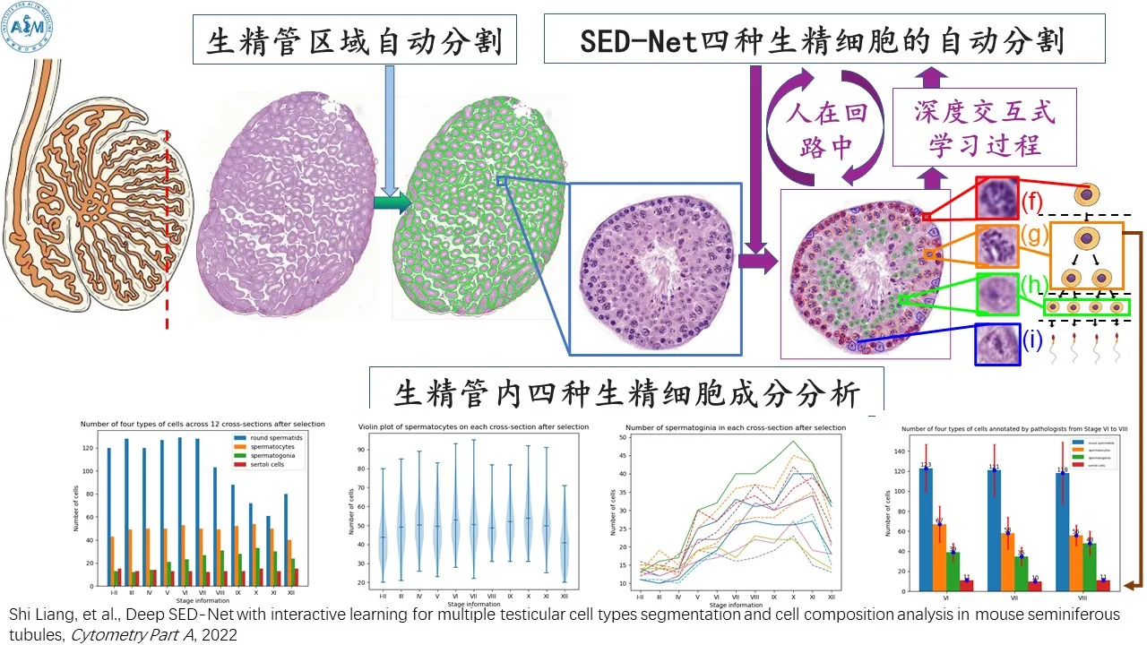
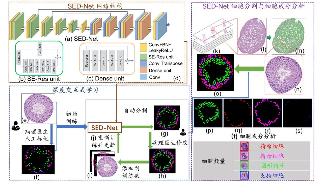
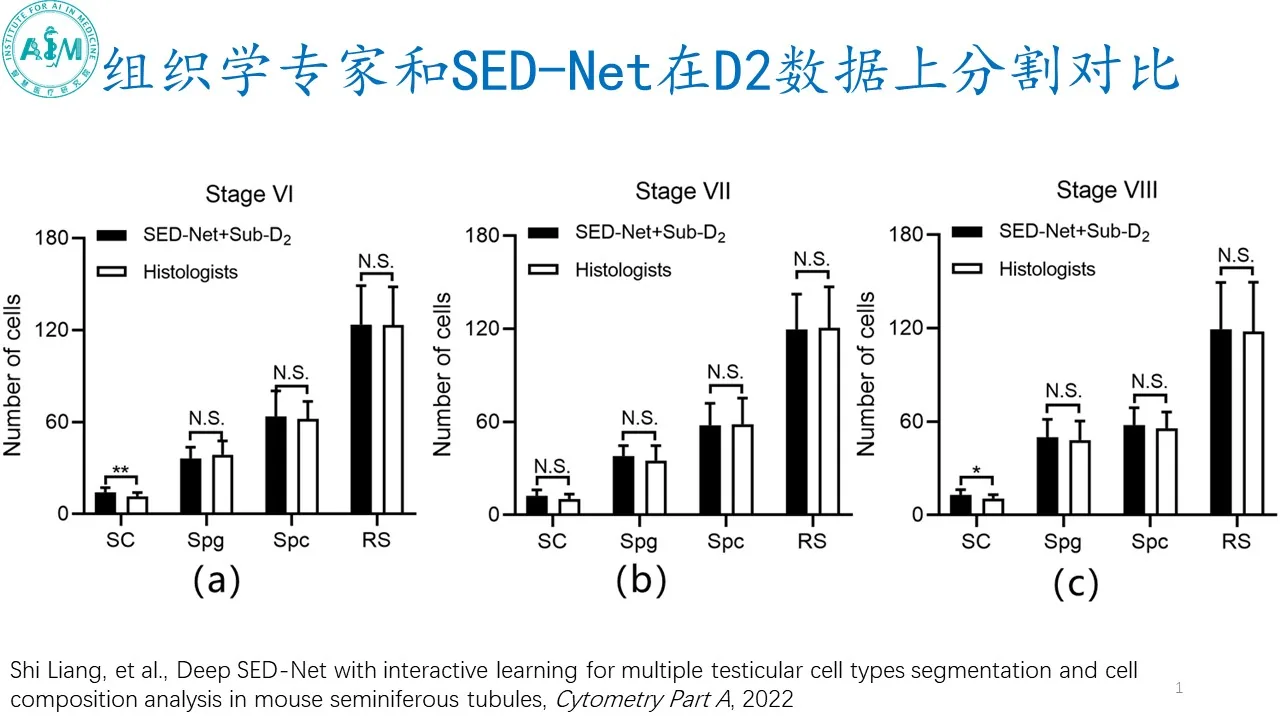
The innovation points of the paper include: 1. Using the idea of “people in the loop”, through the interactive learning mode of histology experts and deep learning models, it is possible to complete the fully automatic segmentation of the four difficult types of spermatogenic cells (spermatogonia, spermatocytes, round spermatozoa, supporting cells) with less annotated data, effectively improving the performance of the model; 2. Establishing a quantitative analysis feature description system for four types of spermatogenic cells in large-scale tissue sections, 3. Quantitatively revealing the presence of seminiferous ducts in I-XII based on quantitative features Change patterns and trends of four types of spermatogenic cells during developmental stages. 4. Aiming at the problem of seminiferous duct analysis, an analysis system composed of multiple deep learning models was established. The four types of spermatogenic cells undergo long and dynamic changes in developmental stages I-XII, and the number of cells that can be artificially labeled is only “a drop in the ocean.” It is extremely challenging to describe this continuous and dynamic change process. In this regard, new analysis frameworks and machine learning models can continue to be explored in the future.
- Haoda Lu, Min Zang, Gabriel Pik Liang Marini, Xiangxue Wang, Yiping Jiao, Nianfei Ao, Kokhaur Ong, Xinmi Huo, Longjie Li, Eugene Yujun Xu, Wilson Wen Bin Goh, Weimiao Yu, Jun Xu , A novel pipeline for computerized mouse spermatogenesis staging , Bioinformatics , 2022.
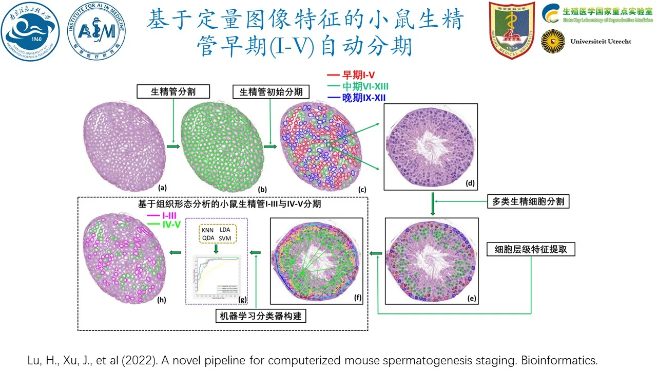
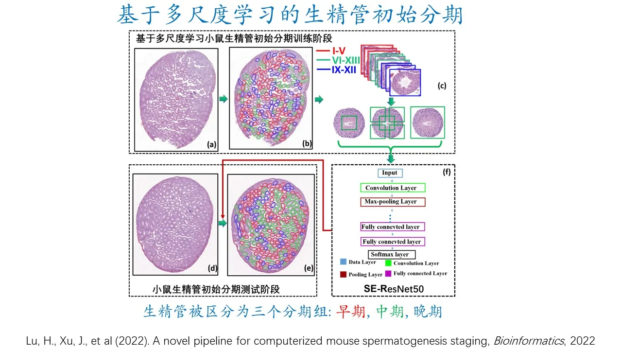
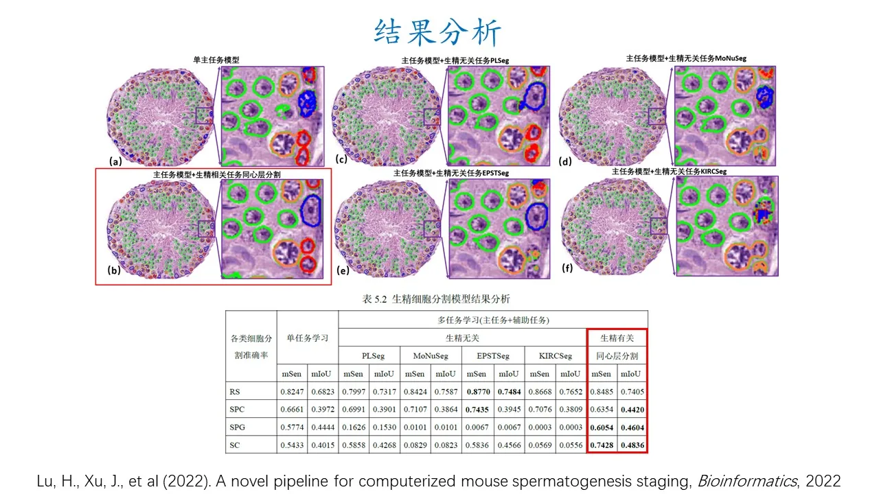
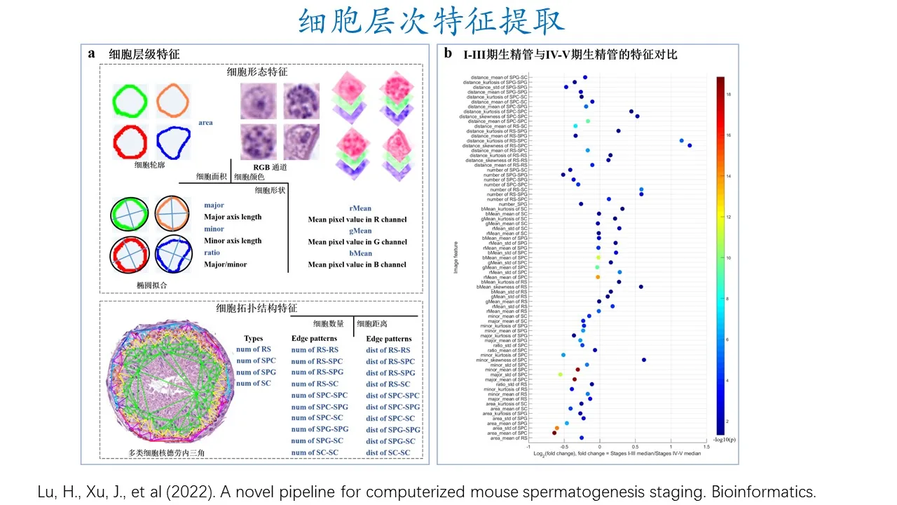
Distinguishing the 12 developmental stages of the mouse seminiferous epithelial cycle is important for understanding the dynamic spermatogenesis process, such as the critical differences between wild-type and infertile mice in stages I-III and IV-V. However, precise differentiation of the stages is extremely challenging due to the morphological similarities between adjacent spermatogenic stages. To this end, this article proposes a novel automated method for the study of spermatogenesis staging (CSS). The CSS model includes four parts: (i) a seminiferous tubule segmentation model for extracting and segmenting each seminiferous tubule; (ii) based on the multi-scale learning (MSL) method to integrate local and global information of the seminiferous tubules to distinguish stages I-V and VI-XII; (iii) based on the multi-task learning (MTL) method to overcome the difficulty of limited training data and achieve fine staging of I-V stages; (iv) established by 204 A description system composed of three-dimensional image features, which distinguishes stage I-III and stage IV-V seminiferous ducts based on cell-level and image-level features. Experimental results show that using the combined MSL and MTL solution proposed in this article, its performance is significantly better than the classic single-scale and single-task models when manual annotation is limited. Furthermore, some image features were found to be distinctive between stages I-III and IV-V, consistent with prior knowledge of pathology. In summary, the CSS model can not only provide a solution for histologists to facilitate quantitative analysis and identification of spermatogenesis stages, but also help them discover new biomarkers from computerized images.
Translated from the original Chinese source.